A systematic review on the definition of periimplantitis: Limits related to the various diagnoses proposed
January 6, 2017 / Categories: Digital Dentistry, Implant Dentistry

Tallarico, Marco

Monje, Alberto

Wang, Hom-Lay

Galindo Moreno, Pablo

Xhanari, Erta

Canullo, Luigi
The aim of this comprehensive systematic review was to present the different definitions of periimplantitis proposed in the literature.
Introduction
The term “periimplantitis” was introduced in the early 1960s to describe infectious pathological conditions of the periimplant tissue.1Levignac J. [Periimplantation osteolysis— periimplantosis—periimplantitis].
→ Rev Fr Odontostomatol. 1965 Oct;12(8):1251–60. French.
Today, periimplantitis is the most frequent complication of dental implants and occurs with a frequency ranging from 1% to 47% at implant level.2Hämmerle CH, Glauser R. Clinical evaluation of dental implant treatment.
→ Periodontol 2000. 2004 Feb;34:230–9.3Schwarz F, Becker K, Sager M. Efficacy of professionally administered plaque removal with or without adjunctive measures for the treatment of peri-implant mucositis. A systematic review and meta-analysis.
→ J Clin Periodontol. 2015 Apr;42 Suppl 16:S202–13.4Atieh MA, Alsabeeha NH, Faggion CM, Duncan WJ. The frequency of peri-implant diseases: a systematic review and meta-analysis.
→ J Periodontol. 2013 Nov;84(11):1586–98.5Zitzmann NU, Margolin MD, Filippi A, Weiger R, Krastl G. Patient assessment and diagnosis in implant treatment.
→ Aust Dent J. 2008 Jun;53 Suppl 1:S3–10.6Sanz M, Chapple IL; Working Group 4 of the VIII European Workshop on Periodontology. Clinical research on peri-implant diseases: consensus report of Working Group 4.
→ J Clin Periodontol. 2012 Feb;39 Suppl 12:202–6.7American Academy of Periodontology. Academy report: peri-implant mucositis and peri-implantitis: a current understanding of their diagnoses and clinical implications.
→ J Periodontol. 2013 Apr;84(4):436–43.8Lang NP, Berglundh T; Working Group 4 of the Seventh European Workshop on Periodontology. Periimplant diseases: where are we now?—Consensus of the Seventh European Workshop on Periodontology.
→ J Clin Periodontol. 2011 Mar;38 Suppl 11:178–81.9Chan HL, Lin GH, Suarez F, MacEachern M, Wang HL. Surgical management of peri-implantitis: a systematic review and meta-analysis of treatment outcomes.
→ J Periodontol. 2014 Aug;85(8):1027–41. Different from periimplant mucositis (defined as the presence of reversible inflammatory soft-tissue infiltrate, without additional bone loss beyond the initial physiological bone remodeling),10Lindhe J, Meyle J; Group D of the European Workshop on Periodontology. Peri-implant diseases: consensus report of the Sixth European Workshop on Periodontology.
→ J Clin Periodontol. 2008 Sep;35(8 Suppl):282–5. periimplantitis has been described as being characterized by an inflammatory process around an implant, including both soft-tissue inflammation and progressive loss of supporting bone beyond the physiological crestal bone remodeling.11Lindhe J, Meyle J; Group D of the European Workshop on Periodontology. Peri-implant diseases: consensus report of the Sixth European Workshop on Periodontology.
→ J Clin Periodontol. 2008 Sep;35(8 Suppl):282–5. However, as highlighted in recent literature reviews and consensus conferences, different definitions of periimplantitis have been reported.12Zitzmann NU, Margolin MD, Filippi A, Weiger R, Krastl G. Patient assessment and diagnosis in implant treatment.
→ Aust Dent J. 2008 Jun;53 Suppl 1:S3–10.13Sanz M, Chapple IL; Working Group 4 of the VIII European Workshop on Periodontology. Clinical research on peri-implant diseases: consensus report of Working Group 4.
→ J Clin Periodontol. 2012 Feb;39 Suppl 12:202–6.14American Academy of Periodontology. Academy report: peri-implant mucositis and peri-implantitis: a current understanding of their diagnoses and clinical implications.
→ J Periodontol. 2013 Apr;84(4):436–43.15Lang NP, Berglundh T; Working Group 4 of the Seventh European Workshop on Periodontology. Periimplant diseases: where are we now?—Consensus of the Seventh European Workshop on Periodontology.
→ J Clin Periodontol. 2011 Mar;38 Suppl 11:178–81.16Mombelli A, Müller N, Cionca N. The epidemiology of peri-implantitis.
→ Clin Oral Implants Res. 2012 Oct;23 Suppl 6:67–76. This may be due in part to the lack of consensus on terminology, etiology, diagnosis and prognosis systems.17Atieh MA, Alsabeeha NH, Faggion CM, Duncan WJ. The frequency of peri-implant diseases: a systematic review and meta-analysis.
→ J Periodontol. 2013 Nov;84(11):1586–98.18Zitzmann NU, Margolin MD, Filippi A, Weiger R, Krastl G. Patient assessment and diagnosis in implant treatment.
→ Aust Dent J. 2008 Jun;53 Suppl 1:S3–10.19Salvi GE, Lang NP. Diagnostic parameters for monitoring peri-implant conditions.
→ Int J Oral Maxillofac Implants. 2004;19 Suppl:116–27.
Periimplantitis has been described as a disease with an infectious component that is similar to chronic periodontitis.20Mombelli A, Van Oosten MA, Schurch EJ, Lang NP. The microbiota associated with successful or failing osseointegrated titanium implants.
→ Oral Microbiol Immunol. 1987 Dec;2(4):145–51. The 8th European Workshop on Periodontology has agreed that the definitions published in 200810 and 20118 should be adopted. The suggested definition should include the following: changes in the level of crestal bone, positive bleeding on probing (BOP) and/or suppuration (SUP), with or without concomitant periimplant pockets (probing pocket depth, PPD).21Lang NP, Berglundh T; Working Group 4 of the Seventh European Workshop on Periodontology. Periimplant diseases: where are we now?—Consensus of the Seventh European Workshop on Periodontology.
→ J Clin Periodontol. 2011 Mar;38 Suppl 11:178–81. Nowadays, although plaque accumulation is still considered the main etiological factor,22Jepsen S, Berglundh T, Genco R, Aass AM, Demirel K, Derks J, Figuero E, Giovannoli JL, Goldstein M, Lambert F, Ortiz-Vigon A, Polyzois I, Salvi GE, Schwarz F, Serino G, Tomasi C, Zitzmann NU. Primary prevention of peri-implantitis: managing peri-implant mucositis.
→ J Clin Periodontol. 2015 Apr;42 Suppl 16:S152–7. it has been shown that there are other potential related risk factors of the disease, including patient, surgical and prosthetic factors that may certainly contribute to its development. 23Albrektsson T, Donos N; Working Group 1. Implant survival and complications. The third EAO consensus conference 2012.
→ Clin Oral Implants Res. 2012 Oct;23 Suppl:63–5.24Heitz-Mayfield LJ. Peri-implant diseases: diagnosis and risk indicators.
→ J Clin Periodontol. 2008 Sep;35(8 Suppl):292–304.25Renvert S, Quirynen M. Risk indicators for peri-implantitis. A narrative review.
→ Clin Oral Implants Res. 2015 Sep;26 Suppl 11:15–44.26Konstantinidis IK, Kotsakis GA, Gerdes S, Walter MH. Cross-sectional study on the prevalence and risk indicators of peri-implant diseases.
→ Eur J Oral Implantol. 2015 Spring;8(1):75–88.27Canullo L, Tallarico M, Radovanovic S, Delibasic B, Covani U, Rakic M. Distinguishing predictive profiles for patient-based risk assessment and diagnostics of plaque induced, surgically and prosthetically triggered peri-implantitis.
→ Clin Oral Implants Res. 2016 Oct;27(10):1243–50. Epub 2015 Nov 20.28Pesce P, Canullo L, Grusovin MG, De Bruyn H, Cosyn J, Pera P. Systematic review of some prosthetic risk factors for periimplantitis.
→ J Prost Dent. 2015 Sep;114(3):346–50.29Canullo L, Schlee M, Wagner W, Covani U; Montegrotto Group for the Study of Peri-implant Disease. International Brainstorming Meeting on Etiologic and Risk Factors of Peri-implantitis, Montegrotto (Padua, Italy), August 2014.
→ Int J Oral Maxillofac Implants. 2015 Sep-Oct;30(5):1093–104.30Albrektsson T, Dahlin C, Jemt T, Sennerby L, Turri A, Wennerberg A. Is marginal bone loss around oral implants the result of a provoked foreign body reaction?
→ Clin Implant Dent Relat Res. 2014 Apr;16(2):155–65.
In the MeSH (Medical Subject Headings) database, the term “periimplantitis” was introduced in 2011 and defined as an inflammatory process with loss of supporting bone in the tissue surrounding functioning dental implants.31MeSH Browser (2011 MeSH) [Internet]. Bethesda (MD): National Library of Medicine (US), Medical Subject Headings Section. [1999] – . Heart failure, systolic; [cited 2011 Jul 8]; [about 1 screen].
→ Available from: http://www.nlm.nih.gov/cgi/mesh/2011/MB_cgi?mode=&index=24306 Despite this very clear and comprehensive disease definition, inconsistencies and confusion emerge in applying the terminology clinically. All of these factors together have led to different interpretations and definitions of this common emerging disease. Besides, recently, the noninfectious foreign-body reaction hypothesis has further complicated the understanding of this issue.32Albrektsson T, Dahlin C, Jemt T, Sennerby L, Turri A, Wennerberg A. Is marginal bone loss around oral implants the result of a provoked foreign body reaction?
→ Clin Implant Dent Relat Res. 2014 Apr;16(2):155–65. The aim of the present systematic review was to present the different definitions of periimplantitis proposed in the literature.
Materials and methods
The present paper was prepared in partial fulfillment of a consensus statement held in Rome, Italy, in January 2016. This systematic review was written according to the PRISMA (Preferred Reporting Items for Systematic Reviews and Meta- Analyses) guidelines (http://www.prismastatement.org/PRISMAStatement/PRISMAStatement.aspx).
The focused question was: Is there an unanimous definition of periimplantitis, including clinical diagnosis. The research question was adapted to the PICO format:
P = population: human patients derived from clinical studies, systematic reviews, narrative reviews, consensuses statements, commentaries or editorials, who presented with at least one dental implant in function for a minimum of one year, affected by periimplantitis;
I = intervention: clinical data collected with the aim of establishing the severity of the periimplant disease and of defining novel criteria by which to classify periimplant diseases;
C = comparator/control: clinical outcomes of periimplantitis compared with clinical signs of periodontitis, as well as with healthy patients;
O = outcomes: clinical parameters and radiographic assessment of periimplantitis: BOP, PPD, bleeding index, presence of SUP and marginal bone loss (MBL).
Search strategy
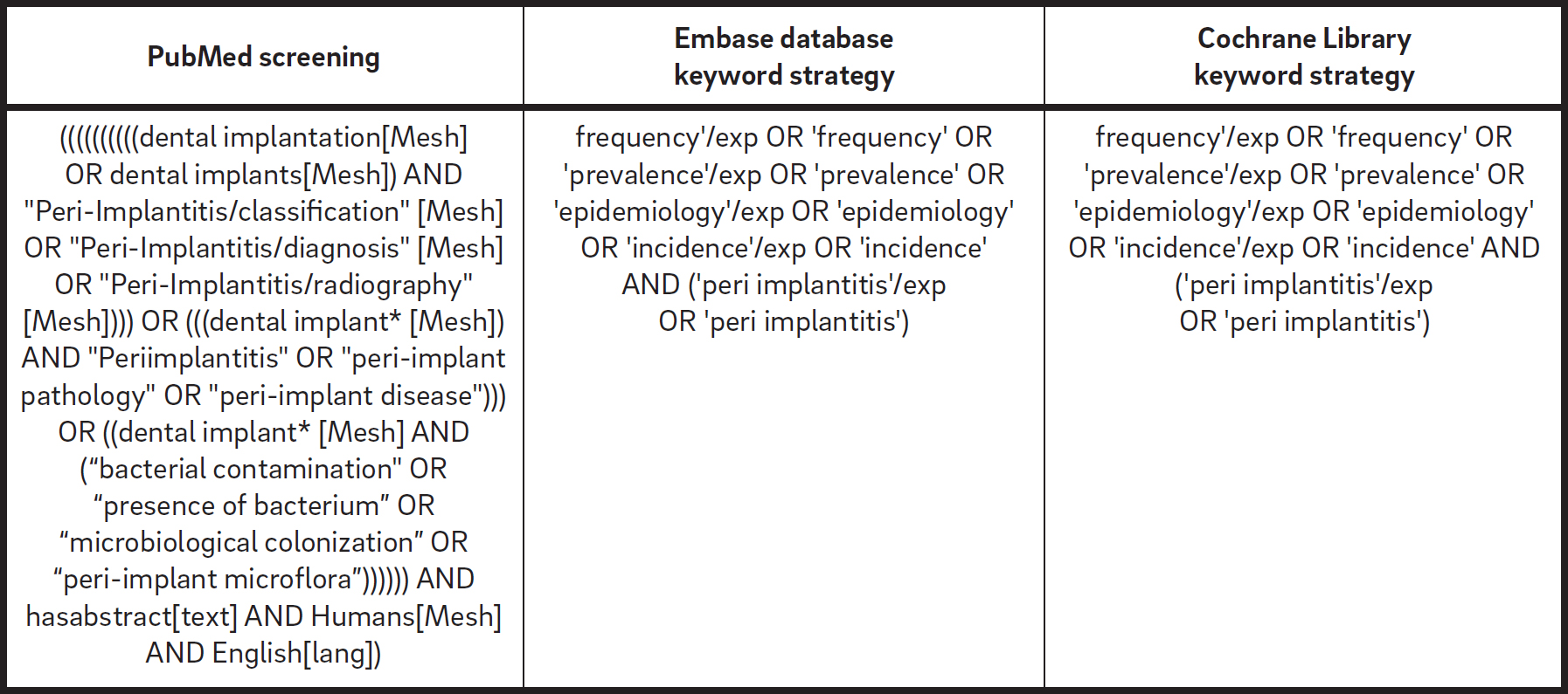
An initial search strategy encompassing the English literature from 1967 up to October 2015 was performed online to identify relevant studies that met the inclusion criteria. The following electronic databases were consulted: PubMed database of the U.S. National Library of Medicine, Embase (Excerpta Medica dataBASE) and the Cochrane Library. According to the AMSTAR (A Measurement Tool to Assess Systematic Reviews) checklist, the Grey Literature Database was screened in the New York Academy of Medicine Grey Literature Report in order to find possible unpublished works. Screening was performed independently and simultaneously by two examiners (MT and AM). A third reviewer (LC) reassessed the included and excluded studies. The electronic databases were searched using a combination of boolean keywords, including MeSH and several free-text terms (Table 1).
Eligibility criteria
The following inclusion criteria were defined for the selection of articles:
- written in English;
- entailing clinical examination of human patients;
- randomized controlled clinical trials of implants of ≥ 1 year in function;
- prospective and retrospective cohort studies of implants of ≥ 1 year in function;
- cross-sectional studies of ≥ 1 year in function; and
- systematic reviews, narrative reviews, consensus statements, commentaries or editorials.
Articles were excluded if they were
- animal studies;
- in vitro studies;
- reports of locally or systemically compromised sites and/or conditions;
- reports with < 15 cases;
- reports involving mini-implants, one-piece implants or blade implants; or
- reports on implants < 1 year in function.
Papers without abstracts, but with titles related to the objectives of this review were selected so that the full text could be screened for eligibility. Full-text papers were obtained for all abstracts and titles that appeared to meet the inclusion criteria and were assessed for inclusion by the same two examiners. The reference lists of the selected studies was screened for additional papers that may have met the eligibility criteria of the study. Additionally, manual searches of the reference lists of selected systematic reviews were conducted, limited to the following journals: Clinical Implant Dentistry and Related Research, Clinical Oral Implants Research, International Journal of Oral and Maxillofacial Implants, Journal of Clinical Periodontology and Journal of Periodontology. Any disagreement between the two reviewers was resolved after an additional discussion. Furthermore, inter-investigator agreement was calculated in the second stage. A final reviewer (LC) evaluated possible inconsistencies between the two reviewers. All of the full texts of the selected papers were stored in shared folders accessible to all of the reviewers.
Qualitative assessment of parameters to define periimplantitis
A descriptive evaluation was performed to analyze qualitatively the range of parameters considered to define periimplantitis as an irreversible inflammatory condition that results in hardtissue breakdown. Accordingly, the following common parameters were appraised: PPD, BOP, SUP and radiographic MBL. Such parameters from the various articles were pooled to analyze the variance or uniformity among the reported case definitions of periimplantitis. Graphs for presenting the variance were generated. While PPD was classified into three different groups (< 3 mm, 3–5 mm and > 5 mm), radiographic MBL was categorized into four main ranges, depen ding on the main reference taking prosthesis delivery as the baseline: ≤ 1 mm, > 1–2 mm, > 3–4 mm and ≥ 5 mm.
Results
Screening process
The combinations of search terms and a manual search of references in selected articles resulted in a list of 1,061 titles. Of these, 976 articles were excluded on the basis of the evaluation of the title and abstract, leaving 85 articles eligible for inclusion (? = 0.84). After application of the eligibility criteria, a total of 49 articles were considered for review. After full-text article selection and reading, the relevant information from each article was extracted. A diagram of the search strategy is shown in Figure 1.

PRISMA flowchart of search strategy.
Definitions of “periimplantitis”
Eighteen manuscripts, including narrative and systematic reviews, consensus statements and original papers, were selected and data were extracted. In 1965, Levignac reported a periimplant soft-tissue inflammation with subsequent destruction of bone and labeled it “periimplantitis.”33Levignac J. [Periimplantation osteolysis— periimplantosis—periimplantitis].
→ Rev Fr Odontostomatol. 1965 Oct;12(8):1251–60. French. In the 1987, Mombelli et al. described periimplantitis as an infectious disease that shares features with chronic periodontitis.34Mombelli A, Van Oosten MA, Schurch EJ, Lang NP. The microbiota associated with successful or failing osseointegrated titanium implants.
→ Oral Microbiol Immunol. 1987 Dec;2(4):145–51. The same author emphasized the infectious nature of this pathological condition, focusing on the bacterial load of the implant surface and the subsequent appearance of a soft-tissue inflammatory reaction adjacent to dental implants that sometimes resulted in loss of supporting bone.35Mombelli A, Müller N, Cionca N. The epidemiology of peri-implantitis.
→ Clin Oral Implants Res. 2012 Oct;23 Suppl 6:67–76.36Mombelli A, Lang NP. The diagnosis and treatment of peri-implantitis.
→ Periodontol 2000. 1998 Jun;17:63–76.37Mombelli A, Décaillet F. The characteristics of biofilms in peri-implant disease.
→ J Clin Periodontol. 2011 Mar;38 Suppl 11:203–13. Like periodontitis, the etiopathogenesis of periimplantitis was shown to be triggered by bacterial infection that activates a cytokine cascade, leading to inflammatory bone loss.38American Academy of Periodontology. Academy report: peri-implant mucositis and peri-implantitis: a current understanding of their diagnoses and clinical implications.
→ J Periodontol. 2013 Apr;84(4):436–43.
“Periimplantitis” became an accepted term in the consensus report from the 1st European Workshop on Periodontology in 1993.39Lang NP. Proceedings of the 1st European Workshop on Periodontology, Charter House at Ittingen, Thurgau, Switzerland, February 1–4, 1993.
→ London: Quintessence; 1994. 478 p. It has been described as an irreversible inflammatory destructive reaction around implants in function that results in loss of supporting bone.40Lang NP. Proceedings of the 1st European Workshop on Periodontology, Charter House at Ittingen, Thurgau, Switzerland, February 1–4, 1993.
→ London: Quintessence; 1994. 478 p. The 6th European Workshop on Periodontology presented a modified definition, not only to acknowledge that periimplantitis is a treatable condition, but also to include the collective term of “periimplant disease” for both periimplant mucositis and periimplantitis.41Lindhe J, Meyle J; Group D of the European Workshop on Periodontology. Peri-implant diseases: consensus report of the Sixth European Workshop on Periodontology.
→ J Clin Periodontol. 2008 Sep;35(8 Suppl):282–5.
In order to improve the quality of research on periimplant diseases, the 7th European Workshop on Periodontology recommended the use of unequivocal case definitions: changes in the level of crestal bone and presence of BOP and/or SUP, with or without concomitant deepening of periimplant pockets.42Lang NP, Berglundh T; Working Group 4 of the Seventh European Workshop on Periodontology. Periimplant diseases: where are we now?—Consensus of the Seventh European Workshop on Periodontology.
→ J Clin Periodontol. 2011 Mar;38 Suppl 11:178–81. Finally, the American Academy of Periodontology in 2013 defined “periimplantitis” as an inflammatory reaction associated with the loss of supporting bone beyond the initial biological bone remodeling around an implant in function.43American Academy of Periodontology. Academy report: peri-implant mucositis and peri-implantitis: a current understanding of their diagnoses and clinical implications.
→ J Periodontol. 2013 Apr;84(4):436–43.
The extent and severity of periimplant diseases have been rarely reported.44Decker AM, Sheridan R, Lin GH, Sutthiboonyapan P, Carroll W, Wang HL. A prognosis system for periimplant diseases.
→ Implant Dent. 2015 Aug;24(4):416–21. 45Froum SJ, Rosen PS. A proposed classification for peri-implantitis.
→ Int J Periodontics Restorative Dent. 2012 Oct;32(5):533–40. Froum and Rosen proposed a combination of BOP and/or SUP, PPD and extent of radiographic MBL around the implant to classify periimplantitis into early, moderate or advanced disease categories.46Froum SJ, Rosen PS. A proposed classification for peri-implantitis.
→ Int J Periodontics Restorative Dent. 2012 Oct;32(5):533–40. Likewise, Decker et al. proposed a prognosis system based on the diagnosis for each category following the Kwok and Caton prognosis classification for natural dentition.47Decker AM, Sheridan R, Lin GH, Sutthiboonyapan P, Carroll W, Wang HL. A prognosis system for periimplant diseases.
→ Implant Dent. 2015 Aug;24(4):416–21. In their study, the authors stated that PPD, extent of radiographic MBL, presence of SUP and implant mobility were found to be the most critical factors for categorizing cases as having a favorable, questionable, unfavorable or hopeless prognosis.48Decker AM, Sheridan R, Lin GH, Sutthiboonyapan P, Carroll W, Wang HL. A prognosis system for periimplant diseases.
→ Implant Dent. 2015 Aug;24(4):416–21.
Recently, Albrektsson et al. modified the concept of periimplantitis as a loss of bone surrounding an implant as a clinically unfavorable, disbalanced foreign-body reaction, specifically stating that osseointegration is a process whereby bone reacts to the dental implant by forming a calcified structure adjacent to it.49Albrektsson T, Dahlin C, Jemt T, Sennerby L, Turri A, Wennerberg A. Is marginal bone loss around oral implants the result of a provoked foreign body reaction?
→ Clin Implant Dent Relat Res. 2014 Apr;16(2):155–65. Indeed, at times, this foreign-body reaction may actually result in osteoclastic activity that may destroy the supporting bone.50Albrektsson T, Dahlin C, Jemt T, Sennerby L, Turri A, Wennerberg A. Is marginal bone loss around oral implants the result of a provoked foreign body reaction?
→ Clin Implant Dent Relat Res. 2014 Apr;16(2):155–65. The authors believe that the term “periimplantitis” is quite appropriate, because it is not a primary disease, but a complication of a clinically unfavorable, disbalanced foreign-body reaction that is the starting point of the pathological process and consequent tissue sequelae.51Albrektsson T, Dahlin C, Jemt T, Sennerby L, Turri A, Wennerberg A. Is marginal bone loss around oral implants the result of a provoked foreign body reaction?
→ Clin Implant Dent Relat Res. 2014 Apr;16(2):155–65.
Currently, as foreseen by the consensus of the 7th European Workshop on Periodontology,52Lang NP, Berglundh T; Working Group 4 of the Seventh European Workshop on Periodontology. Periimplant diseases: where are we now?—Consensus of the Seventh European Workshop on Periodontology.
→ J Clin Periodontol. 2011 Mar;38 Suppl 11:178–81. it is assumed that the infection itself is always caused by plaque and its products; However, numerous risk factors are recognized as being specifically associated with periimplantitis, such as surgical- or prosthetic-related factors,53Canullo L, Tallarico M, Radovanovic S, Delibasic B, Covani U, Rakic M. Distinguishing predictive profiles for patient-based risk assessment and diagnostics of plaque induced, surgically and prosthetically triggered peri-implantitis.
→ Clin Oral Implants Res. 2016 Oct;27(10):1243–50. Epub 2015 Nov 20.54Pesce P, Canullo L, Grusovin MG, De Bruyn H, Cosyn J, Pera P. Systematic review of some prosthetic risk factors for periimplantitis.
→ J Prost Dent. 2015 Sep;114(3):346–50.55Jemt T, Gyzander V, Britse AO. Incidence of surgery related to problems with peri-implantitis: a retrospective study on patients followed up between 2003 and2010 at one specialist clinic.
→ Clin Implant Dent Relat Res. 2015 Apr;17(2):209–20. Epub 2013 Jun 10. implant characteristics,56Canullo L, Schlee M, Wagner W, Covani U; Montegrotto Group for the Study of Peri-implant Disease. International Brainstorming Meeting on Etiologic and Risk Factors of Peri-implantitis, Montegrotto (Padua, Italy), August 2014.
→ Int J Oral Maxillofac Implants. 2015 Sep-Oct;30(5):1093–104. smoking57Sgolastra F, Petrucci A, Severino M, Gatto R, Monaco A. Smoking and the risk of peri-implantitis. A systematic review and meta-analysis.
→ Clin Oral Impl Res. 2015 Apr;26(4):e62–7. and host response.58Sgolastra F, Petrucci A, Severino M, Gatto R, Monaco A. Smoking and the risk of peri-implantitis. A systematic review and meta-analysis.
→ Clin Oral Impl Res. 2015 Apr;26(4):e62–7. 59Schwarz F, Becker K, Sahm N, Horstkemper T, Rousi K, Becker J. The prevalence of peri-implant diseases for two-piece implants with an internal tube-in-tube connection: a cross-sectional analysis of 512 implants.
→ Clin Oral Implants Res. 2015 Jul 14. doi:10.1111/clr.12609. [Epub ahead of print]
Definition of periimplantitis with clinical and radiographic diagnosis
Thirty-one manuscripts (Table 2) were selected and data were extracted. Informations from 1,711 patients with 5,432 implants were analyzed. The term “periimplantitis” has generally been used to describe any implant with varying degrees of bone loss, and a clear definition was either not presented or was extracted directly from the terminology.
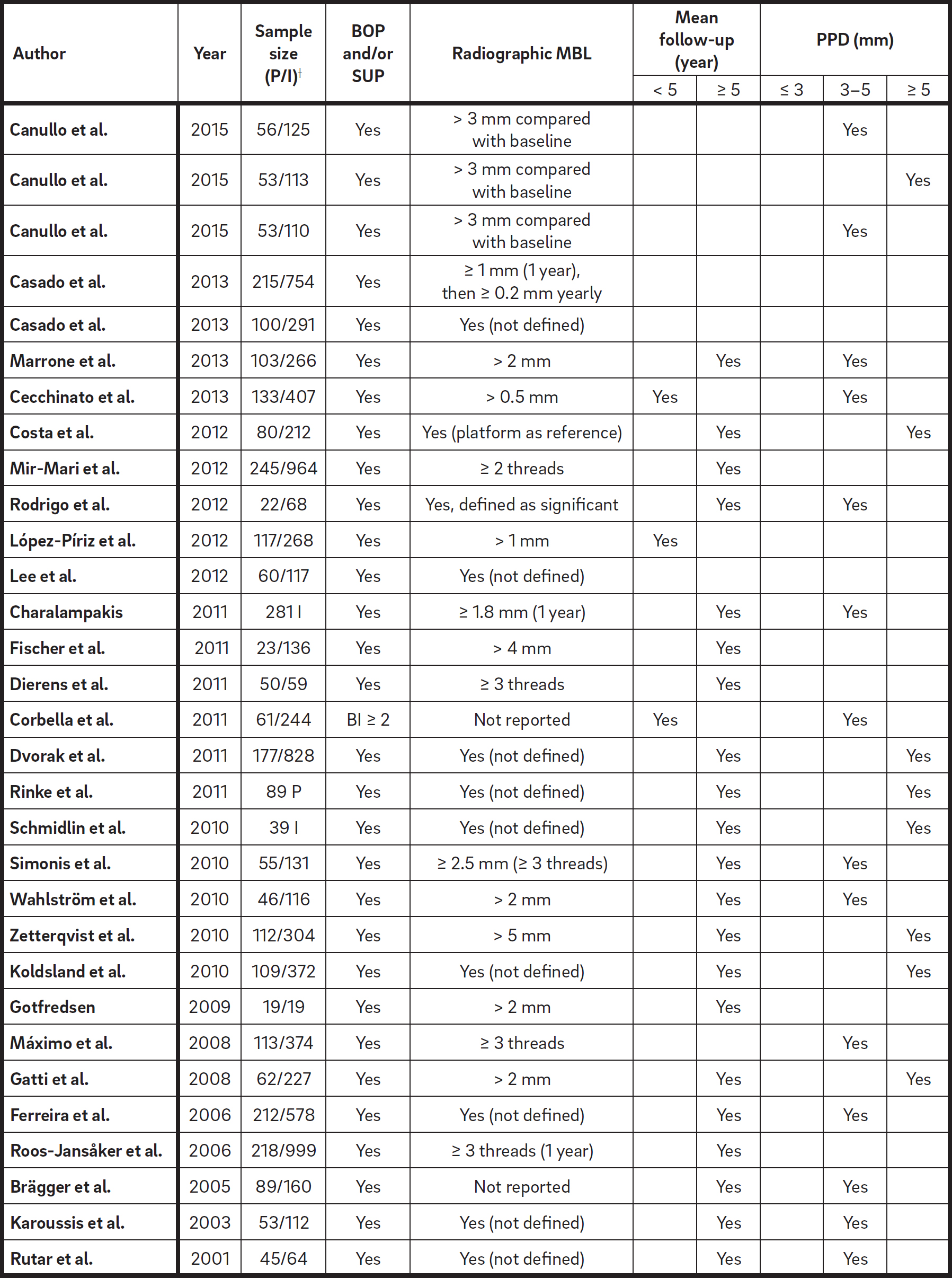
Four main characteristics have been used to define “periimplantitis”. Interestingly, all of the authors consider BOP and SUP as indicators of periimplantitis. This approach considers purely plaque- and foreign-body-induced periimplantitis, where an inflammatory response is often triggered by the biofilm or its products and/ or foreign substances, such as residual cement. Moreover, 22 studies clearly reported PPD as a crucial parameter for determining periimplantitis. No study considered PPD < 3 mm as indicative of periimplantitis. While the vast majority (64%) of the studies defined PPD = 3–5 mm as indicative of periimplantitis, the remaining 36% considered PPD > 5 mm as the reference (Fig. 2). A radiographic MBL ≥ 0.5–1 mm, > 1–2 mm, > 3–4 mm and ≥ 5 mm, taking prosthesis delivery as baseline, was considered as defining periimplantitis in 15%, 45%, 35% and 5% of the studies, respectively (Fig. 3). As such, it was speculated that a radiographic MBL < 1 mm should be considered as physiological bone remodeling.
BOP and/or SUP were prerequisite in all of the analyzed studies. In most of the studies, the combination of clinical and radiographic measurements were used for case definition. In two prospective studies,60Corbella S, Del Fabbro M, Taschieri S, De Siena F, Francetti L. Clinical evaluation of an implant maintenance protocol for the prevention of peri-implant diseases in patients treated with immediately loaded full-arch rehabilitations.
→ Int J Dent Hyg. 2011 Aug;9(3):216–22.61Brägger U, Karoussis I, Persson R, Pjetursson B, Salvi G, Lang N. Technical and biological complications/failures with single crowns and fixed partial dentures on implants: a 10-year prospective cohort study.
→ Clin Oral Implants Res. 2005 Jun;16(3):326–34. the radiographic MBL were not reported, and clinical measurements alone were used to assess biological complications. In these cases, the presence of BOP and/or SUP on probing and PPD ≥ 4 mm were prerequisite for a diagnosis of periimplantitis. In nine studies, one randomized controlled trial,62Fischer K, Stenberg T. Prospective 10-year cohort study based on a randomized, controlled trial (RCT) on implant-supported full-arch maxillary prostheses. part II: prosthetic outcomes and maintenance.
→ Clin Implant Dent Relat Res. 2013 Aug;15(4):498–508. three prospective 63Casado PL, Pereira MC, Duarte MEL, Granjeiro JM. History of chronic periodontitis is a high risk indicator for peri-implant disease.
→ Braz Dent J. 2013 Mar-Apr;24(2):136–41. 64Mir-Mari J, Mir-Orfila P, Figueiredo R, Valmaseda-Castellón E, Gay-Escoda C. Prevalence of peri-implant diseases. A cross-sectional study based on a private practice environment.
→ J Clin Periodontol. 2012 May;39(5):490–494. Epub 2012 April 9.65Gotfredsen K. A 10-year prospective study of single tooth implants placed in the anterior maxilla.
→ Clin Implant Dent Relat Res. 2012 Mar;14(1):80–7. Epub 2009 Aug 6. and five retrospective studies,66López-Píriz R, Morales A, Giménez MJ, Bowen A, Carroquino R, Aguilar L, Corral I, del Val C, González I, Ilzarbe LM, Maestre JR, Padullés E, Torres-Lear F, Granizo JJ, San-Román F, Hernández S, Prieto J; SEIRN group. Correlation between clinical parameters characterising peri-implant and periodontal health: a practice-based research in Spain in a series of patients with implants installed 4-5 years ago.
→ Med Oral Patol Oral Cir Bucal. 2012 Sep 1;17(5):e893–901. 67Cecchinato D, Parpaiola A, Lindhe J. A cross-sectional study on the prevalence of marginal bone loss among implant patients.
→ Clin Oral Implants Res. 2013 Jan;24(1):87–90. 68Casado PL, Villas-Boas R, de Mello W, Duarte ME, Granjeiro JM. Peri-implant disease and chronic periodontitis: is interleukin-6 gene promoter polymorphism the common risk factor in a Brazilian population?
→ Int J Oral Maxillofac Implants. 2013 Jan-Feb;28(1):35–43.69Cho-Yan Lee J, Mattheos N, Nixon KC, Ivanovski S. Residual periodontal pockets are a risk indicator for peri-implantitis in patients treated for periodontitis.
→ Clin Oral Implants Res. 2012 Mar;23(3):325–33. 70Dierens M, Vandeweghe S, Kisch J, Nilner K, De Bruyn H. Long-term follow-up of turned single implants placed in periodontally healthy patients after 16–22 years: radiographic and peri-implant outcome.
→ Clin Oral Implants Res. 2012 Feb;23(2):197–204. Epub 2011 Jul 6. BOP and radiographic assessments were performed alone, without reporting any PPD measurements. In these cases, a MBL ranging from 0.5 mm39 to > 4 mm34 was considered to be associated with periimplantitis. Before 2012, changes in the level of crestal bone were either not defined or not clearly reported, making the diagnosis of periimplantitis difficult.71Sanz M, Chapple IL; Working Group 4 of the VIII European Workshop on Periodontology. Clinical research on peri-implant diseases: consensus report of Working Group 4.
→ J Clin Periodontol. 2012 Feb;39 Suppl 12:202–6.72Lang NP, Berglundh T; Working Group 4 of the Seventh European Workshop on Periodontology. Periimplant diseases: where are we now?—Consensus of the Seventh European Workshop on Periodontology.
→ J Clin Periodontol. 2011 Mar;38 Suppl 11:178–81.73Corbella S, Del Fabbro M, Taschieri S, De Siena F, Francetti L. Clinical evaluation of an implant maintenance protocol for the prevention of peri-implant diseases in patients treated with immediately loaded full-arch rehabilitations.
→ Int J Dent Hyg. 2011 Aug;9(3):216–22.74Brägger U, Karoussis I, Persson R, Pjetursson B, Salvi G, Lang N. Technical and biological complications/failures with single crowns and fixed partial dentures on implants: a 10-year prospective cohort study.
→ Clin Oral Implants Res. 2005 Jun;16(3):326–34.75Cho-Yan Lee J, Mattheos N, Nixon KC, Ivanovski S. Residual periodontal pockets are a risk indicator for peri-implantitis in patients treated for periodontitis.
→ Clin Oral Implants Res. 2012 Mar;23(3):325–33.76Dvorak G, Arnhart C, Heuberer S, Huber CD, Watzek G, Gruber R. Peri-implantitis and late implant failures in postmenopausalwomen: a cross-sectional study.
→ J Clin Periodontol. 2011 Oct;38(10):950–5.77Rinke S, Ohl S, Ziebolz D, Lange K, Eickholz P. Prevalence of periimplant disease in partially edentulous patients: a practice-based cross-sectional study.
→ Clin Oral Implants Res. 2011 Aug;22(8):826–33.78Schmidlin K, Schnell N, Steiner S, Salvi GE, Pjetursson B, Matuliene G, Zwahlen M, Brägger U, Lang NP. Complication and failure rates in patients treated for chronic periodontitis and restored with single crowns on teeth and/or implants.
→ Clin Oral Implants Res. 2010 May;21(5):550–7.79Ferreira SD, Silva GL, Cortelli JR, Costa JE, Costa FO. Prevalence and risk variables for peri-implant disease in Brazilian subjects.
→ J Clin Periodontol. 2006 Dec;33(12):929–35.80Koldsland OC, Scheie AA, Aass AM. Prevalence of peri-implantitis related to severity of the disease with different degrees of bone loss.
→ J Periodontol. 2010 Feb;81(2):231–8. 81Karoussis IK, Salvi GE, Heitz-Mayfield LJ, Brägger U, Hämmerle CH, Lang NP. Long-term implant prognosis in patients with and without a history of chronic periodontitis: a 10-year prospective cohort study of the ITI Dental Implant System.
→ Clin Oral Implants Res. 2003 Jun;14(3):329–39. 82Rutar A, Lang NP, Buser D, Burgin W, Mombelli A. Retrospective assessment of clinical and microbiological factors affecting periimplant tissue conditions.
→ Clin Oral Implants Res. 2001 Jun;12(3):189–95. However, even in studies that defined the entity of MBL, different diagnostic criteria were used. In one long-term study, periimplantitis was defined as the presence of BOP, PPD ≥ 4 mm and MBL > 0.5 mm.39 How ever, another study used MBL > 4 mm as a reference value.83Fischer K, Stenberg T. Prospective 10-year cohort study based on a randomized, controlled trial (RCT) on implant-supported full-arch maxillary prostheses. part II: prosthetic outcomes and maintenance.
→ Clin Implant Dent Relat Res. 2013 Aug;15(4):498–508. Most of the studies considered MBL > 2 mm for the diagnosis of periimplantitis. 84Mir-Mari J, Mir-Orfila P, Figueiredo R, Valmaseda-Castellón E, Gay-Escoda C. Prevalence of peri-implant diseases. A cross-sectional study based on a private practice environment.
→ J Clin Periodontol. 2012 May;39(5):490–494. Epub 2012 April 9.85Rodrigo D, Martin C, Sanz M. Biological complications and peri-implant clinical and radiographic changes at immediately placed dental implants. A prospective 5-year cohort study.
→ Clin Oral Implants Res. 2012 Oct;23(10):1224–31.86Marrone A, Lasserre J, Bercy P, Brecx MC. Prevalence and risk factors for peri-implant disease in Belgian adults.
→ Clin Oral Implants Res. 2013 Aug;24(8):934–40.87Gotfredsen K, Wiskott A; Working Group 4. Consensus report—reconstructions on implants. The third EAO Consensus Conference 2012.
→ Clin Oral Implants Res. 2012 Oct;23 Suppl 6:238–41. 88Gatti C, Gatti F, Chiapasco M, Esposito M. Outcome of dental implants in partially edentulous patients with and without a history of periodontitis: a 5-year interim analysis of a cohort study.
→ Eur J Oral Implantol. 2008 Spring;1(1):45–51.89Wahlström M, Sagulin GB, Jansson LE. Clinical follow-up of unilateral, fixed dental prosthesis on maxillary implants.
→ Clin Oral Implants Res. 2010 Nov;21(11):1294–300. Previously, our group used a radiographic MBL > 3 mm, from the baseline radiograph taken at the time of prosthesis delivery, to diagnose periimplantitis.90Canullo L, Tallarico M, Radovanovic S, Delibasic B, Covani U, Rakic M. Distinguishing predictive profiles for patient-based risk assessment and diagnostics of plaque induced, surgically and prosthetically triggered peri-implantitis.
→ Clin Oral Implants Res. 2016 Oct;27(10):1243–50. Epub 2015 Nov 20.91Canullo L, Peñarrocha-Oltra D, Covani U, Botticelli D, Serino G, Peñarrocha M. Clinical and microbiological findings in patients with peri-implantitis: a cross-sectional study.
→ Clin Oral Implants Res. 2016 Mar;27(3):376–82. Epub 2015 Jan 26.92Canullo L, Peñarrocha D, Covani U, Rossetti PH. Microbiologic and clinical findings of implants in healthy condition and with peri-implantitis.
→ Int J Oral Maxillofac Implants. 2015 Jul-Aug;30(4):834–42. In three other studies, MBL was considered in relation to the time that the prosthesis was in function.93Casado PL, Pereira MC, Duarte MEL, Granjeiro JM. History of chronic periodontitis is a high risk indicator for peri-implant disease.
→ Braz Dent J. 2013 Mar-Apr;24(2):136–41.94Charalampakis G, Leonhardt A, Rabe P, Dahlén G. Clinical and microbiological characteristics of peri-implantitis cases: a retrospective multicentre study.
→ Clin Oral Implants Res. 2012 Sep;23(9):1045–54.95Roos-Jansåker AM, Lindahl C, Renvert H, Renvert S. Nine- to fourteen-year follow-up of implant treatment. Part II: presence of peri-implant lesions.
→ J Clin Periodontol. 2006 Apr;33(4):290–5. All of the studies but five calculated MBL in millimeters. In the other studies, the implant threads were used as reference.96Mir-Mari J, Mir-Orfila P, Figueiredo R, Valmaseda-Castellón E, Gay-Escoda C. Prevalence of peri-implant diseases. A cross-sectional study based on a private practice environment.
→ J Clin Periodontol. 2012 May;39(5):490–494. Epub 2012 April 9.97Dierens M, Vandeweghe S, Kisch J, Nilner K, De Bruyn H. Long-term follow-up of turned single implants placed in periodontally healthy patients after 16–22 years: radiographic and peri-implant outcome.
→ Clin Oral Implants Res. 2012 Feb;23(2):197–204. Epub 2011 Jul 6.98Roos-Jansåker AM, Lindahl C, Renvert H, Renvert S. Nine- to fourteen-year follow-up of implant treatment. Part II: presence of peri-implant lesions.
→ J Clin Periodontol. 2006 Apr;33(4):290–5.99Simonis P, Dufour T, Tenenbaum H.Long-term implant survival and success: a 10-16-year follow-up of non-submerged dental implants.
→ Clin Oral Implants Res. 2010 Jul;21(7):772–7.100Máximo MB, de Mendonça AC, Alves JF, Cortelli SC, Peruzzo DC, Duarte PM. Peri-implant diseases may be associated with increased time loading and generalized periodontal bone loss:preliminary results.
→ J Oral Implantol. 2008 Oct;34(5):268–73. Eight studies applied PPD > 5 mm for the diagnosis of periimplantitis.101Dvorak G, Arnhart C, Heuberer S, Huber CD, Watzek G, Gruber R. Peri-implantitis and late implant failures in postmenopausalwomen: a cross-sectional study.
→ J Clin Periodontol. 2011 Oct;38(10):950–5.102Rinke S, Ohl S, Ziebolz D, Lange K, Eickholz P. Prevalence of periimplant disease in partially edentulous patients: a practice-based cross-sectional study.
→ Clin Oral Implants Res. 2011 Aug;22(8):826–33.103Koldsland OC, Scheie AA, Aass AM. Prevalence of peri-implantitis related to severity of the disease with different degrees of bone loss.
→ J Periodontol. 2010 Feb;81(2):231–8. 104Gatti C, Gatti F, Chiapasco M, Esposito M. Outcome of dental implants in partially edentulous patients with and without a history of periodontitis: a 5-year interim analysis of a cohort study.
→ Eur J Oral Implantol. 2008 Spring;1(1):45–51.105Simonis P, Dufour T, Tenenbaum H.Long-term implant survival and success: a 10-16-year follow-up of non-submerged dental implants.
→ Clin Oral Implants Res. 2010 Jul;21(7):772–7.106Canullo L, Peñarrocha-Oltra D, Covani U, Rossetti P. Microbiologic and clinical findings of implants in healthy condition and with peri-implantitis.
→ Int J Oral Maxillofac Implants. 2015 Jul-Aug;30(4):834–42.107Zetterqvist L, Feldman S, Rotter B, Vincenzi G, Wennström JL, Chierico A, Stach RM, Kenealy JN. A prospective, multicenter, randomized-controlled 5-year study of hybrid and fully etched implants for the incidence of peri-implantitis.
→ J Periodontol. 2010 Apr;81(4):493–501. 108Costa FO, Takenaka-Martinez S, Cota LO, Ferreira SD, Silva GL, Costa JE. Peri-implant disease in subjects with and without preventive maintenance: a 5-year follow-up.
→ J Clin Periodontol. 2012 Feb;39(2):173–81. ) Marrone et al. defined periimplantitis as the presence of BOP, PPD > 5 mm and MBL > 2 mm.109Marrone A, Lasserre J, Bercy P, Brecx MC. Prevalence and risk factors for peri-implant disease in Belgian adults.
→ Clin Oral Implants Res. 2013 Aug;24(8):934–40. Charalampakis et al. applied the criteria of the presence of BOP and/or SUP, PPD ≥ 5 mm and MBL ≥ 1.8 mm after one year in function.110Charalampakis G, Leonhardt A, Rabe P, Dahlén G. Clinical and microbiological characteristics of peri-implantitis cases: a retrospective multicentre study.
→ Clin Oral Implants Res. 2012 Sep;23(9):1045–54. Zetterqvist et al. included cases of PPD > 5 mm and MBL ≥ 3 mm.111Zetterqvist L, Feldman S, Rotter B, Vincenzi G, Wennström JL, Chierico A, Stach RM, Kenealy JN. A prospective, multicenter, randomized-controlled 5-year study of hybrid and fully etched implants for the incidence of peri-implantitis.
→ J Periodontol. 2010 Apr;81(4):493–501. Two other studies, one prospective112Costa FO, Takenaka-Martinez S, Cota LO, Ferreira SD, Silva GL, Costa JE. Peri-implant disease in subjects with and without preventive maintenance: a 5-year follow-up.
→ J Clin Periodontol. 2012 Feb;39(2):173–81. and one retrospective,113Dvorak G, Arnhart C, Schön P, Heuberer S, Watzek G, Gahleitner A. The “puffed cheek method” to evaluate mucosal thickness: case series.
→ Clin Oral Implants Res. 2012 Jul;24(7):719–24. applied the presence of BOP and/or SUP, PPD > 5 mm and radiographic signs of MBL, without specifying the baseline bone level. Positive BOP and/or SUP, radiographic MBL ≥ 3 mm and PPD ≥ 6 mm were used by Koldsland et al.114Koldsland OC, Scheie AA, Aass AM. Prevalence of peri-implantitis related to severity of the disease with different degrees of bone loss.
→ J Periodontol. 2010 Feb;81(2):231–8.
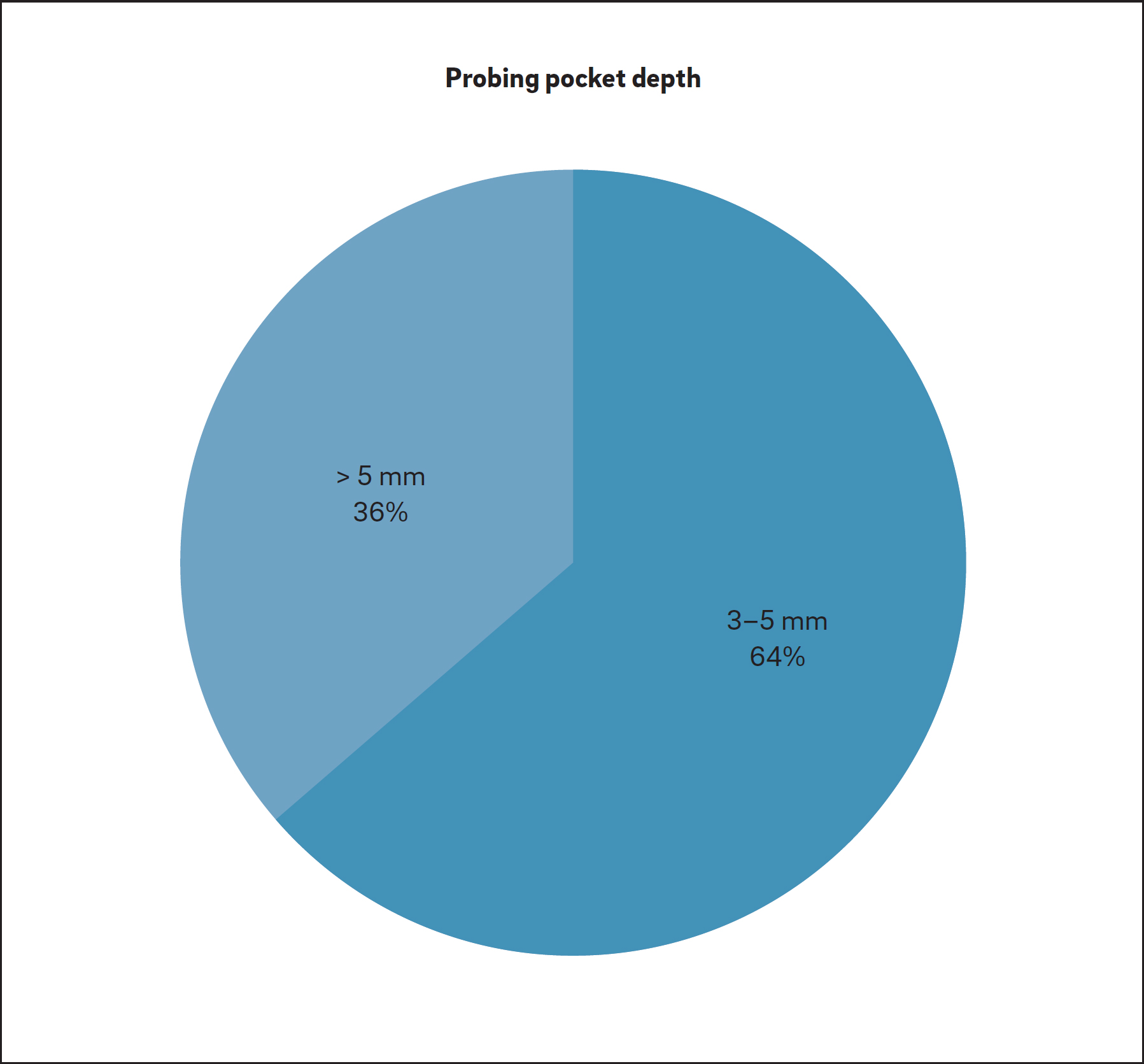
Percentage of the included studies relating to different PPD ranges used to define periimplantitis.

Percentage of the included studies relating to different radiographic MBL values used to define periimplantitis.
At the 7th and 8th European Workshop on Periodontology, periimplant mucositis and periimplantitis were described as follows: “changes in the level of crestal bone, presence of bleeding on probing and/or suppuration; with or without concomitant deepening of peri-implant pockets.”115Sanz M, Chapple IL; Working Group 4 of the VIII European Workshop on Periodontology. Clinical research on peri-implant diseases: consensus report of Working Group 4.
→ J Clin Periodontol. 2012 Feb;39 Suppl 12:202–6.116Lang NP, Berglundh T; Working Group 4 of the Seventh European Workshop on Periodontology. Periimplant diseases: where are we now?—Consensus of the Seventh European Workshop on Periodontology.
→ J Clin Periodontol. 2011 Mar;38 Suppl 11:178–81. Periimplant mucositis was defined with positive BOP and/or SUP and periimplantitis with positive BOP and/or SUP, in combination with radiographic MBL ≥ 2 mm. The same parameters were used by Zitzmann and Berglundh to define periimplantitis. 117Zitzmann NU, Berglundh T. Definition and prevalence of peri-implant diseases.
→ J Clin Periodontol.2008 Sep;35(8 Suppl):286–91. However, Atieh et al. used the same criteria, plus PPD ≥ 5 mm, as the definition of periimplantitis in their systematic review paper.118Atieh MA, Alsabeeha NH, Faggion CM, Duncan WJ. The frequency of peri-implant diseases: a systematic review and meta-analysis.
→ J Periodontol. 2013 Nov;84(11):1586–98.
Discussion
Periimplant diseases present in two forms: periimplant mucositis and periimplantitis.119Lindhe J, Meyle J; Group D of the European Workshop on Periodontology. Peri-implant diseases: consensus report of the Sixth European Workshop on Periodontology.
→ J Clin Periodontol. 2008 Sep;35(8 Suppl):282–5. Both are characterized by an inflammatory reaction in the tissue surrounding an implant. Periimplant mucositis has been defined as a reversible inflammatory reaction in the soft-tissue surrounding an implant in function, whereas periimplantitis has been defined as a more profound inflammatory lesion characterized by a deepened periimplant pocket and loss of supporting bone around a functional implant.120Lindhe J, Meyle J; Group D of the European Workshop on Periodontology. Peri-implant diseases: consensus report of the Sixth European Workshop on Periodontology.
→ J Clin Periodontol. 2008 Sep;35(8 Suppl):282–5.121Mombelli A, Lang NP. The diagnosis and treatment of peri-implantitis.
→ Periodontol 2000. 1998 Jun;17:63–76.
Studies published in early 2010 suggested that mucositis and periimplantitis are equivalent to periodontitis, since both are described as an imbalance between bacterial load and the host response.122Lang NP, Berglundh T; Working Group 4 of the Seventh European Workshop on Periodontology. Periimplant diseases: where are we now?—Consensus of the Seventh European Workshop on Periodontology.
→ J Clin Periodontol. 2011 Mar;38 Suppl 11:178–81.123Mombelli A, Décaillet F. The characteristics of biofilms in peri-implant disease.
→ J Clin Periodontol. 2011 Mar;38 Suppl 11:203–13. Based upon this, both diseases are closely related to the formation of a biofilm containing microbiota rich in Gram-negative bacteria in the presence of a susceptible host.124Heitz-Mayfield LJ, Lang NP. Comparative biology of chronic and aggressive periodontitis vs. peri-implantitis.
→ Periodontol 2000. 2010 Jun;53:167–81. However, it has been shown that microorganisms may be present, but are not a necessity for periimplantitis.125Heitz-Mayfield LJ, Lang NP. Comparative biology of chronic and aggressive periodontitis vs. peri-implantitis.
→ Periodontol 2000. 2010 Jun;53:167–81. In addition, both periodontitis and periimplantitis share several common systemic risk factors or indicators (e.g., smoking, poor oral hygiene, diabetes or history of periodontitis, osteoporosis). 126Lindhe J, Meyle J; Group D of the European Workshop on Periodontology. Peri-implant diseases: consensus report of the Sixth European Workshop on Periodontology.
→ J Clin Periodontol. 2008 Sep;35(8 Suppl):282–5.127Dvorak G, Arnhart C, Schön P, Heuberer S, Watzek G, Gahleitner A. The “puffed cheek method” to evaluate mucosal thickness: case series.
→ Clin Oral Implants Res. 2012 Jul;24(7):719–24.128Heitz-Mayfield LJ, Lang NP. Comparative biology of chronic and aggressive periodontitis vs. peri-implantitis.
→ Periodontol 2000. 2010 Jun;53:167–81.129Tamura N, Ochi M, Miyakawa H, Nakazawa F. Analysis of bacterial flora associated with peri-implantitis using obligate anaerobic culture technique and 16S rDNA gene sequence.
→ Int J Oral Maxillofac Implants. 2013 Nov-Dec;28(6):1521–9. Similarly, periimplantitis, as occurs with periodontitis, seems to be influenced by a particular genetic profile (i.e., interleukin-1 polymorphism).130Liao J, Li C, Wang Y, Ten M, Sun X, Tian A, Zhang Q, Liang X. Meta-analysis of the association between common interleukin-1 polymorphisms and dental implant failure.
→ Mol Biol Rep. 2014 May;41(5):2789–98. Others have rejected the description of a disease comparable to periodontitis,131Albrektsson T, Dahlin C, Jemt T, Sennerby L, Turri A, Wennerberg A. Is marginal bone loss around oral implants the result of a provoked foreign body reaction?
→ Clin Implant Dent Relat Res. 2014 Apr;16(2):155–65.132Mouhyi J, Dohan Ehrenfest DM, Albrektsson T. The peri-implantitis: implant surfaces, microstructure, and physicochemical aspects.
→ Clin Implant Dent Relat Res. 2012 Apr;14(2):170–83. Epub 2009 Oct 16.133Becker ST, Beck-Broichsitter BE, Graetz C, Dörfer CE, Wiltfang J, Häsler R. Peri-Implantitis versus periodontitis: functional differences indicated by transcriptome profiling.
→ Clin Implant Dent Relat Res. 2014 Jun;16(3):401–11. Epub 2012 Sep 11. because of the anatomical differences that exist between periodontal and periimplant structures (e.g., different collagen fiber orientation [perpendicular vs. horizontal], vascularity or repair capacity, and the mechanical resilience provided by the periodontal ligament).134Berglundh T, Lindhe J. Dimension of the periimplant mucosa. Biological width revisited.
→ J Clin Periodontol. 1996 Oct;23(10):971–3. In fact, periodontitis is characterized by inflammatory destruction of the supporting apparatus of the dentition (periodontium), including the periodontal ligament and alveolar bone. Owing to the different composition of the two supporting tissues, similar tissue reactions around an implant and a tooth seem most unlikely. The term “osseoinsufficiency” was proposed by Zarb and Koka to describe the difference between periimplantitis and periodontitis-induced bone loss.135Zarb GA, Koka S. Osseointegration: promise and platitudes.
→ Int J Prosthodont. 2012 Jan-Feb;25(1):11–2. The anatomical image of bone resorption due to periodontitis or periimplantitis differs, in many situations with very wide bone craters being typical for the implant but not for the tooth. Hence, periimplantitis may be considered distinct from periodontitis in that it significantly differs regarding onset and progression and has poor treatment predictability; consequently, its treatment must be focused on early diagnosis and controlling the risk factors or indicators to prevent it from occurring.
To date, there have been no standardized parameters to clinically differentiate the various stages and severities of periimplantitis.136Froum SJ, Rosen PS. A proposed classification for peri-implantitis.
→ Int J Periodontics Restorative Dent. 2012 Oct;32(5):533–40. The criteria used to diagnose periimplantitis remain inconclusive. Most existing studies used clinical parameters in combination with radiographic findings to define periimplantitis. However, clinical parameters such as BOP and PPD around implants are less predictable, since they are influenced by more confounding factors compared with natural dentition.137Hämmerle CH, Glauser R. Clinical evaluation of dental implant treatment.
→ Periodontol 2000. 2004 Feb;34:230–9.138Schwarz F, Becker K, Sager M. Efficacy of professionally administered plaque removal with or without adjunctive measures for the treatment of peri-implant mucositis. A systematic review and meta-analysis.
→ J Clin Periodontol. 2015 Apr;42 Suppl 16:S202–13. Furthermore, any factor that facilitates plaque formation (e.g., poor oral hygiene) or host defense capability (e.g., smoking habit, excessive alcohol consumption, genetic traits, history of periodontitis or use of bisphosphonates) might contribute to the development of periimplantitis.139Heitz-Mayfield LJ. Peri-implant diseases: diagnosis and risk indicators.
→ J Clin Periodontol. 2008 Sep;35(8 Suppl):292–304.140Renvert S, Aghazadeh A, Hallström H, Persson GR. Factors related to periimplantitis— a retrospective study.
→ Clin Oral Implants Res. 2014 Apr;25(4):522–9.141Pjetursson BE, Thoma D, Jung RE, Zwahlen M, Zembic A. A systematic review of the survival and complication rates of implant-supported fixed dental prostheses (FDPs) after a mean observation period of at least 5 years.
→ Clin Oral Implants Res. 2012 Oct;23 Suppl 6:22–38. The diagnosis and progression of periimplantitis may be characterized by increased measurements for clinical parameters (PPD, BOP, SUP or even mobility), MBL and microbiology. Regarding clinical parameters, PPD is a valid method of assessment, as correlation exists between bone levels recorded and radiographic probe penetration.142Cho-Yan Lee J, Mattheos N, Nixon KC, Ivanovski S. Residual periodontal pockets are a risk indicator for peri-implantitis in patients treated for periodontitis.
→ Clin Oral Implants Res. 2012 Mar;23(3):325–33.143Quirynen M, van Steenberghe D, Jacobs R, Schotte A, Darius P. The reliability of pocket probing around screw-type implants.
→ Clin Oral Implants Res. 1991 Oct-Dec;2(4):186–92. Nevertheless, in a cross-sectional study, the intraoperatively measured periimplant bone levels were more apical than the radiographic bone levels.144García-García M, Mir-Marí J, Benic GI, Figueiredo R,Valmaseda-Castellón E. Accuracy of periapical radiography in assessing bone level in implants affected by peri-implantitis: a cross-sectional study.
→ J Clinic Periodontol. 2015. 2016 Jan;43(1):85–91. Epub 2016 Feb 12. SUP occurs more frequently in implants with than without progressive bone loss, particularly in smokers, and may be associated with episodes of active tissue destruction.145Atieh MA, Alsabeeha NH, Faggion CM, Duncan WJ. The frequency of peri-implant diseases: a systematic review and meta-analysis.
→ J Periodontol. 2013 Nov;84(11):1586–98. In a systematic review, Berglundh et al. defined periimplantitis as having a PPD ≥ 6 mm or MBL ≥ 2.5 mm.146Berglundh T, Persson L, Klinge B. A systematic review of the incidence of biological and technical complications in implant dentistry reported in prospective longitudinal studies of at least 5 years.
→ J Clin Periodontol. 2002 Dec;29 Suppl 3:197–212; discussion 232–3. Lang and Berglundh, in the 2011 European Federation of Periodontology consensus, stated that clinical and radiographic data should routinely be obtained after prosthesis installation on implants in order to establish a baseline for the diagnosis of periimplantitis during maintenance of implant patients. 147Lang NP, Berglundh T; Working Group 4 of the Seventh European Workshop on Periodontology. Periimplant diseases: where are we now?—Consensus of the Seventh European Workshop on Periodontology.
→ J Clin Periodontol. 2011 Mar;38 Suppl 11:178–81. A meta-analysis by Derks and Tomasi clear ly showed a positive relationship between the prevalence of periimplantitis and function time.148Derks J, Tomasi C. Peri-implant health and disease. A systematic review of current epidemiology.
→ J Clin Periodontol. 2015 Apr;42 Suppl 16:S158–71. The presence of bone loss and PPD alone may not be enough to establish a diagnosis of periimplantitis.149Derks J, Tomasi C. Peri-implant health and disease. A systematic review of current epidemiology.
→ J Clin Periodontol. 2015 Apr;42 Suppl 16:S158–71. One important factor that potentially influences the wide range of periimplantitis prevalence is the lack of consensus regarding the clinical parameters. For example, one study reported that if PPD > 4 mm was used as criterion, then 74.8% individuals had periimplantitis, but if this measurement was changed to > 6 mm, then the prevalence dropped to 43.9%.150Koldsland OC, Scheie AA, Aass AM. Prevalence of peri-implantitis related to severity of the disease with different degrees of bone loss.
→ J Periodontol. 2010 Feb;81(2):231–8. When radiographic MBL was considered for defining periimplantitis, 25.3% individuals showed > 2 mm, while 13.1% had > 3 mm.151Koldsland OC, Scheie AA, Aass AM. Prevalence of peri-implantitis related to severity of the disease with different degrees of bone loss.
→ J Periodontol. 2010 Feb;81(2):231–8. Indeed, if PPD is considered, some further heterogeneity can be found. Probing around implants is influenced by many factors, such as the size of the probe, the probing force, the direction of the probe, the health and thickness of the periimplant softtissue, and the design of the implant neck and the superstructure.152Levignac J. [Periimplantation osteolysis— periimplantosis—periimplantitis].
→ Rev Fr Odontostomatol. 1965 Oct;12(8):1251–60. French. In fact, the platform-switched design, as well as defective restorations, can complicate probing and, thus, hide the true extent of periimplantitis.153Heitz-Mayfield LJ. Peri-implant diseases: diagnosis and risk indicators.
→ J Clin Periodontol. 2008 Sep;35(8 Suppl):292–304.154Renvert S, Quirynen M. Risk indicators for peri-implantitis. A narrative review.
→ Clin Oral Implants Res. 2015 Sep;26 Suppl 11:15–44.155Heitz-Mayfield L, Mombelli A. The therapy of peri-implantitis: a systematic review.
→ Int J Oral Maxillofac Implants.2014;29 Suppl:325–45. Furthermore, the pre sence of discrepancies in the buccolingual hard- and soft-tissue levels may result in different PPD readings.
Owing to the lack of standard parameters to determine the presence and severity of periimplant disease, it is difficult to develop a clinical strategy based upon PPD in managing this common problem in implant dentistry. However, Froum and Rosen proposed a classification system to determine periimplantitis severity based upon PPD, MBL and clinical signs of BOP and/or SUP,156Froum SJ, Rosen PS. A proposed classification for peri-implantitis.
→ Int J Periodontics Restorative Dent. 2012 Oct;32(5):533–40. but this system remains to be validated. Furthermore, in a series of studies by Merli et al., the inter-rater agreement in the diagnosis of periimplant disease was judged as merely good, owing to the unclear definition of periimplantitis and mucositis, with complete agreement obtained only in half of the cases (52%).157Halazonetis TD, Haffajee AD, Socransky SS. Relationship of clinical parameters to attachment loss in subsets of subjects with destructive periodontal diseases.
→ J Clin Periodontol. 1989 Oct;16(9):563–8.
The vast majority (45%) of the studies included in the present review found radiographic MBL > 1–2 mm after prosthetic loading. Hence, the following criteria for defining periimplantitis are proposed: a radiographic MBL > 1 mm after implant prosthesis delivery or 2 mm at least six months after implant prosthesis placement as a good indicator of periimplantitis. BOP does not possess a high predictive value owing to the weak soft-tissue connection around dental implants. Likewise, PPD largely relies on implant design (bone vs. tissue level), apicocoronal position and biotype. From the extracted data, it seems logical to consider radiographic MBL as the most uniform and accurate indicator of periimplantitis. Although, the cut-off value depends on the patient’s inflammatory pattern, the type of surgery, the apicocoronal implant position, the implant’s macrodesign and the crestal module, considering the rapid disease progression over time, strict radiographic control must be followed if any clinical symptom is detected. Furthermore, the clinician must use a combination of the many available clinical parameters, such as PPD, inflammatory status of the mucosa, BOP on light probing, radiographic MBL, and possibly bacterial and/or periimplant crevicular fluid biomarkers to establish an accurate diagnosis of periimplantitis.158Froum SJ, Rosen PS. A proposed classification for peri-implantitis.
→ Int J Periodontics Restorative Dent. 2012 Oct;32(5):533–40. Unlike in the case of periodontitis, bacterial testing may not reliable in diagnosing periimplantitis.159Dabdoub SM, Tsigarida AA, Kumar PS. Patient-specific analysis of periodontal and peri-implant microbiomes.
→ J Dent Res. 2013 Dec;92(12 Suppl):168S–75S. This suggests that periodontal and periimplant ecosystems differ significantly and, hence, periimplant disease might not always be approached as an infectious disease. Similarly, such difference has been shown to apply to the pathogenesis.160Carcuac O, Berglundh T. Composition of human peri-implantitis and periodontitislesions.
→ J Dent Res. 2014 Nov;93(11):1083–8. Furthermore, no evidence was found that primary infection caused marginal bone resorption.((Qian J, Wennerberg A, Albrektsson T. Reasons for marginal bone loss around oral implants.
→ Clin Implant Dent Relat Res. 2012 Dec;14(6):792–807.
Conclusion
The available scientific literature suggested an absence of a unanimous definition of periimplantitis. Actual definitions of periimplantitis were based solely on clinical parameters without consideration of other potential related risk factors of the disease. Future studies that apply consistent case definitions should be considered.
The authors declare no conflict of interests.
Acknowledgments
The authors wish to thank Dr. Mia Rakic for her scientific contribution to this work.
Interview
with Tallarico Marco
Why did you conduct the research reported on in this paper?
The present paper was prepared in partial fulfillment of a consensus statement held in Rome, Italy, in January 2016, with the aim of presenting the various definitions of periimplantitis proposed in the literature.
For what reasons could others cite your paper?
The present paper concluded that definition of periimplantitis was based on clinical parameters and therefore not supported by the current available evidence. Furthermore, clinicians could cite the present manuscript regarding its finding that there is an absence of a unanimous definition on periimplantitis in the current literature.
How could your study’s findings have an impact on dentistry?
The definition of periimplantitis based on clinical parameters is not supported by current literature. Furthermore, clinicians have to consider that multiple risk factors could act synergistically in the particular clinical scenario, making identification of the leading risk factors more difficult.
What is the relevance of your study’s findings to the daily practice of a dentist?
Dentists should not consider periimplantitis as a purely bacterial disease; they should rather consider and prevent any potential risk factors that may result in periimplant bone loss.
What are your recommendations for further investigation of the topic of your article?
With regard to the implications for future clinical research, quality assessment of reporting on human studies on diagnostic criteria for periimplantitis should be performed to accurately describe periimplant disease. Longitudinal findings that consider surgical-, prosthetic- and patient-related factors, as well as an evidence-based, etiology-driven classification, should also be performed, to better guide the clinician in clinical decision-making to prevent or treat periimplantitis.
References
1, 33. ↑ Levignac J. [Periimplantation osteolysis— periimplantosis—periimplantitis].
→ Rev Fr Odontostomatol. 1965 Oct;12(8):1251–60. French. 2. ↑ Hämmerle CH, Glauser R. Clinical evaluation of dental implant treatment.
→ Periodontol 2000. 2004 Feb;34:230–9. 3. ↑ Schwarz F, Becker K, Sager M. Efficacy of professionally administered plaque removal with or without adjunctive measures for the treatment of peri-implant mucositis. A systematic review and meta-analysis.
→ J Clin Periodontol. 2015 Apr;42 Suppl 16:S202–13. 4, 17. ↑ Atieh MA, Alsabeeha NH, Faggion CM, Duncan WJ. The frequency of peri-implant diseases: a systematic review and meta-analysis.
→ J Periodontol. 2013 Nov;84(11):1586–98. 5, 12, 18. ↑ Zitzmann NU, Margolin MD, Filippi A, Weiger R, Krastl G. Patient assessment and diagnosis in implant treatment.
→ Aust Dent J. 2008 Jun;53 Suppl 1:S3–10. 6, 13, 71. ↑ Sanz M, Chapple IL; Working Group 4 of the VIII European Workshop on Periodontology. Clinical research on peri-implant diseases: consensus report of Working Group 4.
→ J Clin Periodontol. 2012 Feb;39 Suppl 12:202–6. 7, 14, 38, 43. ↑ American Academy of Periodontology. Academy report: peri-implant mucositis and peri-implantitis: a current understanding of their diagnoses and clinical implications.
→ J Periodontol. 2013 Apr;84(4):436–43. 8, 15, 21, 42, 52, 72. ↑ Lang NP, Berglundh T; Working Group 4 of the Seventh European Workshop on Periodontology. Periimplant diseases: where are we now?—Consensus of the Seventh European Workshop on Periodontology.
→ J Clin Periodontol. 2011 Mar;38 Suppl 11:178–81. 9. ↑ Chan HL, Lin GH, Suarez F, MacEachern M, Wang HL. Surgical management of peri-implantitis: a systematic review and meta-analysis of treatment outcomes.
→ J Periodontol. 2014 Aug;85(8):1027–41. 10, 11, 41. ↑ Lindhe J, Meyle J; Group D of the European Workshop on Periodontology. Peri-implant diseases: consensus report of the Sixth European Workshop on Periodontology.
→ J Clin Periodontol. 2008 Sep;35(8 Suppl):282–5. 16, 35. ↑ Mombelli A, Müller N, Cionca N. The epidemiology of peri-implantitis.
→ Clin Oral Implants Res. 2012 Oct;23 Suppl 6:67–76. 19. ↑ Salvi GE, Lang NP. Diagnostic parameters for monitoring peri-implant conditions.
→ Int J Oral Maxillofac Implants. 2004;19 Suppl:116–27. 20, 34. ↑ Mombelli A, Van Oosten MA, Schurch EJ, Lang NP. The microbiota associated with successful or failing osseointegrated titanium implants.
→ Oral Microbiol Immunol. 1987 Dec;2(4):145–51. 22. ↑ Jepsen S, Berglundh T, Genco R, Aass AM, Demirel K, Derks J, Figuero E, Giovannoli JL, Goldstein M, Lambert F, Ortiz-Vigon A, Polyzois I, Salvi GE, Schwarz F, Serino G, Tomasi C, Zitzmann NU. Primary prevention of peri-implantitis: managing peri-implant mucositis.
→ J Clin Periodontol. 2015 Apr;42 Suppl 16:S152–7. 23. ↑ Albrektsson T, Donos N; Working Group 1. Implant survival and complications. The third EAO consensus conference 2012.
→ Clin Oral Implants Res. 2012 Oct;23 Suppl:63–5. 24. ↑ Heitz-Mayfield LJ. Peri-implant diseases: diagnosis and risk indicators.
→ J Clin Periodontol. 2008 Sep;35(8 Suppl):292–304. 25. ↑ Renvert S, Quirynen M. Risk indicators for peri-implantitis. A narrative review.
→ Clin Oral Implants Res. 2015 Sep;26 Suppl 11:15–44. 26. ↑ Konstantinidis IK, Kotsakis GA, Gerdes S, Walter MH. Cross-sectional study on the prevalence and risk indicators of peri-implant diseases.
→ Eur J Oral Implantol. 2015 Spring;8(1):75–88. 27, 53, 90. ↑ Canullo L, Tallarico M, Radovanovic S, Delibasic B, Covani U, Rakic M. Distinguishing predictive profiles for patient-based risk assessment and diagnostics of plaque induced, surgically and prosthetically triggered peri-implantitis.
→ Clin Oral Implants Res. 2016 Oct;27(10):1243–50. Epub 2015 Nov 20. 28, 54. ↑ Pesce P, Canullo L, Grusovin MG, De Bruyn H, Cosyn J, Pera P. Systematic review of some prosthetic risk factors for periimplantitis.
→ J Prost Dent. 2015 Sep;114(3):346–50. 29, 56. ↑ Canullo L, Schlee M, Wagner W, Covani U; Montegrotto Group for the Study of Peri-implant Disease. International Brainstorming Meeting on Etiologic and Risk Factors of Peri-implantitis, Montegrotto (Padua, Italy), August 2014.
→ Int J Oral Maxillofac Implants. 2015 Sep-Oct;30(5):1093–104. 30, 32, 49, 50, 51. ↑ Albrektsson T, Dahlin C, Jemt T, Sennerby L, Turri A, Wennerberg A. Is marginal bone loss around oral implants the result of a provoked foreign body reaction?
→ Clin Implant Dent Relat Res. 2014 Apr;16(2):155–65. 31. ↑ MeSH Browser (2011 MeSH) [Internet]. Bethesda (MD): National Library of Medicine (US), Medical Subject Headings Section. [1999] – . Heart failure, systolic; [cited 2011 Jul 8]; [about 1 screen].
→ Available from: http://www.nlm.nih.gov/cgi/mesh/2011/MB_cgi?mode=&index=24306 36. ↑ Mombelli A, Lang NP. The diagnosis and treatment of peri-implantitis.
→ Periodontol 2000. 1998 Jun;17:63–76. 37. ↑ Mombelli A, Décaillet F. The characteristics of biofilms in peri-implant disease.
→ J Clin Periodontol. 2011 Mar;38 Suppl 11:203–13. 39, 40. ↑ Lang NP. Proceedings of the 1st European Workshop on Periodontology, Charter House at Ittingen, Thurgau, Switzerland, February 1–4, 1993.
→ London: Quintessence; 1994. 478 p. 44, 47, 48. ↑ Decker AM, Sheridan R, Lin GH, Sutthiboonyapan P, Carroll W, Wang HL. A prognosis system for periimplant diseases.
→ Implant Dent. 2015 Aug;24(4):416–21. 45, 46. ↑ Froum SJ, Rosen PS. A proposed classification for peri-implantitis.
→ Int J Periodontics Restorative Dent. 2012 Oct;32(5):533–40. 55. ↑ Jemt T, Gyzander V, Britse AO. Incidence of surgery related to problems with peri-implantitis: a retrospective study on patients followed up between 2003 and2010 at one specialist clinic.
→ Clin Implant Dent Relat Res. 2015 Apr;17(2):209–20. Epub 2013 Jun 10. 57, 58. ↑ Sgolastra F, Petrucci A, Severino M, Gatto R, Monaco A. Smoking and the risk of peri-implantitis. A systematic review and meta-analysis.
→ Clin Oral Impl Res. 2015 Apr;26(4):e62–7. 59. ↑ Schwarz F, Becker K, Sahm N, Horstkemper T, Rousi K, Becker J. The prevalence of peri-implant diseases for two-piece implants with an internal tube-in-tube connection: a cross-sectional analysis of 512 implants.
→ Clin Oral Implants Res. 2015 Jul 14. doi:10.1111/clr.12609. [Epub ahead of print] 60. ↑ Corbella S, Del Fabbro M, Taschieri S, De Siena F, Francetti L. Clinical evaluation of an implant maintenance protocol for the prevention of peri-implant diseases in patients treated with immediately loaded full-arch rehabilitations.
→ Int J Dent Hyg. 2011 Aug;9(3):216–22. 61. ↑ Brägger U, Karoussis I, Persson R, Pjetursson B, Salvi G, Lang N. Technical and biological complications/failures with single crowns and fixed partial dentures on implants: a 10-year prospective cohort study.
→ Clin Oral Implants Res. 2005 Jun;16(3):326–34. 62. ↑ Fischer K, Stenberg T. Prospective 10-year cohort study based on a randomized, controlled trial (RCT) on implant-supported full-arch maxillary prostheses. part II: prosthetic outcomes and maintenance.
→ Clin Implant Dent Relat Res. 2013 Aug;15(4):498–508. 63. ↑ Casado PL, Pereira MC, Duarte MEL, Granjeiro JM. History of chronic periodontitis is a high risk indicator for peri-implant disease.
→ Braz Dent J. 2013 Mar-Apr;24(2):136–41. 64. ↑ Mir-Mari J, Mir-Orfila P, Figueiredo R, Valmaseda-Castellón E, Gay-Escoda C. Prevalence of peri-implant diseases. A cross-sectional study based on a private practice environment.
→ J Clin Periodontol. 2012 May;39(5):490–494. Epub 2012 April 9. 65. ↑ Gotfredsen K. A 10-year prospective study of single tooth implants placed in the anterior maxilla.
→ Clin Implant Dent Relat Res. 2012 Mar;14(1):80–7. Epub 2009 Aug 6. 66. ↑ López-Píriz R, Morales A, Giménez MJ, Bowen A, Carroquino R, Aguilar L, Corral I, del Val C, González I, Ilzarbe LM, Maestre JR, Padullés E, Torres-Lear F, Granizo JJ, San-Román F, Hernández S, Prieto J; SEIRN group. Correlation between clinical parameters characterising peri-implant and periodontal health: a practice-based research in Spain in a series of patients with implants installed 4-5 years ago.
→ Med Oral Patol Oral Cir Bucal. 2012 Sep 1;17(5):e893–901. 67. ↑ Cecchinato D, Parpaiola A, Lindhe J. A cross-sectional study on the prevalence of marginal bone loss among implant patients.
→ Clin Oral Implants Res. 2013 Jan;24(1):87–90. 68. ↑ Casado PL, Villas-Boas R, de Mello W, Duarte ME, Granjeiro JM. Peri-implant disease and chronic periodontitis: is interleukin-6 gene promoter polymorphism the common risk factor in a Brazilian population?
→ Int J Oral Maxillofac Implants. 2013 Jan-Feb;28(1):35–43. 69. ↑ Cho-Yan Lee J, Mattheos N, Nixon KC, Ivanovski S. Residual periodontal pockets are a risk indicator for peri-implantitis in patients treated for periodontitis.
→ Clin Oral Implants Res. 2012 Mar;23(3):325–33. 70. ↑ Dierens M, Vandeweghe S, Kisch J, Nilner K, De Bruyn H. Long-term follow-up of turned single implants placed in periodontally healthy patients after 16–22 years: radiographic and peri-implant outcome.
→ Clin Oral Implants Res. 2012 Feb;23(2):197–204. Epub 2011 Jul 6. 73. ↑ Corbella S, Del Fabbro M, Taschieri S, De Siena F, Francetti L. Clinical evaluation of an implant maintenance protocol for the prevention of peri-implant diseases in patients treated with immediately loaded full-arch rehabilitations.
→ Int J Dent Hyg. 2011 Aug;9(3):216–22. 74. ↑ Brägger U, Karoussis I, Persson R, Pjetursson B, Salvi G, Lang N. Technical and biological complications/failures with single crowns and fixed partial dentures on implants: a 10-year prospective cohort study.
→ Clin Oral Implants Res. 2005 Jun;16(3):326–34. 75. ↑ Cho-Yan Lee J, Mattheos N, Nixon KC, Ivanovski S. Residual periodontal pockets are a risk indicator for peri-implantitis in patients treated for periodontitis.
→ Clin Oral Implants Res. 2012 Mar;23(3):325–33. 76, 101. ↑ Dvorak G, Arnhart C, Heuberer S, Huber CD, Watzek G, Gruber R. Peri-implantitis and late implant failures in postmenopausalwomen: a cross-sectional study.
→ J Clin Periodontol. 2011 Oct;38(10):950–5. 77, 102. ↑ Rinke S, Ohl S, Ziebolz D, Lange K, Eickholz P. Prevalence of periimplant disease in partially edentulous patients: a practice-based cross-sectional study.
→ Clin Oral Implants Res. 2011 Aug;22(8):826–33. 78. ↑ Schmidlin K, Schnell N, Steiner S, Salvi GE, Pjetursson B, Matuliene G, Zwahlen M, Brägger U, Lang NP. Complication and failure rates in patients treated for chronic periodontitis and restored with single crowns on teeth and/or implants.
→ Clin Oral Implants Res. 2010 May;21(5):550–7. 79. ↑ Ferreira SD, Silva GL, Cortelli JR, Costa JE, Costa FO. Prevalence and risk variables for peri-implant disease in Brazilian subjects.
→ J Clin Periodontol. 2006 Dec;33(12):929–35. 80, 103. ↑ Koldsland OC, Scheie AA, Aass AM. Prevalence of peri-implantitis related to severity of the disease with different degrees of bone loss.
→ J Periodontol. 2010 Feb;81(2):231–8. 81. ↑ Karoussis IK, Salvi GE, Heitz-Mayfield LJ, Brägger U, Hämmerle CH, Lang NP. Long-term implant prognosis in patients with and without a history of chronic periodontitis: a 10-year prospective cohort study of the ITI Dental Implant System.
→ Clin Oral Implants Res. 2003 Jun;14(3):329–39. 82. ↑ Rutar A, Lang NP, Buser D, Burgin W, Mombelli A. Retrospective assessment of clinical and microbiological factors affecting periimplant tissue conditions.
→ Clin Oral Implants Res. 2001 Jun;12(3):189–95. 83. ↑ Fischer K, Stenberg T. Prospective 10-year cohort study based on a randomized, controlled trial (RCT) on implant-supported full-arch maxillary prostheses. part II: prosthetic outcomes and maintenance.
→ Clin Implant Dent Relat Res. 2013 Aug;15(4):498–508. 84, 96. ↑ Mir-Mari J, Mir-Orfila P, Figueiredo R, Valmaseda-Castellón E, Gay-Escoda C. Prevalence of peri-implant diseases. A cross-sectional study based on a private practice environment.
→ J Clin Periodontol. 2012 May;39(5):490–494. Epub 2012 April 9. 85. ↑ Rodrigo D, Martin C, Sanz M. Biological complications and peri-implant clinical and radiographic changes at immediately placed dental implants. A prospective 5-year cohort study.
→ Clin Oral Implants Res. 2012 Oct;23(10):1224–31. 86. ↑ Marrone A, Lasserre J, Bercy P, Brecx MC. Prevalence and risk factors for peri-implant disease in Belgian adults.
→ Clin Oral Implants Res. 2013 Aug;24(8):934–40. 87. ↑ Gotfredsen K, Wiskott A; Working Group 4. Consensus report—reconstructions on implants. The third EAO Consensus Conference 2012.
→ Clin Oral Implants Res. 2012 Oct;23 Suppl 6:238–41. 88, 104. ↑ Gatti C, Gatti F, Chiapasco M, Esposito M. Outcome of dental implants in partially edentulous patients with and without a history of periodontitis: a 5-year interim analysis of a cohort study.
→ Eur J Oral Implantol. 2008 Spring;1(1):45–51. 89. ↑ Wahlström M, Sagulin GB, Jansson LE. Clinical follow-up of unilateral, fixed dental prosthesis on maxillary implants.
→ Clin Oral Implants Res. 2010 Nov;21(11):1294–300. 91. ↑ Canullo L, Peñarrocha-Oltra D, Covani U, Botticelli D, Serino G, Peñarrocha M. Clinical and microbiological findings in patients with peri-implantitis: a cross-sectional study.
→ Clin Oral Implants Res. 2016 Mar;27(3):376–82. Epub 2015 Jan 26. 92. ↑ Canullo L, Peñarrocha D, Covani U, Rossetti PH. Microbiologic and clinical findings of implants in healthy condition and with peri-implantitis.
→ Int J Oral Maxillofac Implants. 2015 Jul-Aug;30(4):834–42. 93. ↑ Casado PL, Pereira MC, Duarte MEL, Granjeiro JM. History of chronic periodontitis is a high risk indicator for peri-implant disease.
→ Braz Dent J. 2013 Mar-Apr;24(2):136–41. 94. ↑ Charalampakis G, Leonhardt A, Rabe P, Dahlén G. Clinical and microbiological characteristics of peri-implantitis cases: a retrospective multicentre study.
→ Clin Oral Implants Res. 2012 Sep;23(9):1045–54. 95, 98. ↑ Roos-Jansåker AM, Lindahl C, Renvert H, Renvert S. Nine- to fourteen-year follow-up of implant treatment. Part II: presence of peri-implant lesions.
→ J Clin Periodontol. 2006 Apr;33(4):290–5. 97. ↑ Dierens M, Vandeweghe S, Kisch J, Nilner K, De Bruyn H. Long-term follow-up of turned single implants placed in periodontally healthy patients after 16–22 years: radiographic and peri-implant outcome.
→ Clin Oral Implants Res. 2012 Feb;23(2):197–204. Epub 2011 Jul 6. 99, 105. ↑ Simonis P, Dufour T, Tenenbaum H.Long-term implant survival and success: a 10-16-year follow-up of non-submerged dental implants.
→ Clin Oral Implants Res. 2010 Jul;21(7):772–7. 100. ↑ Máximo MB, de Mendonça AC, Alves JF, Cortelli SC, Peruzzo DC, Duarte PM. Peri-implant diseases may be associated with increased time loading and generalized periodontal bone loss:preliminary results.
→ J Oral Implantol. 2008 Oct;34(5):268–73. 106. ↑ Canullo L, Peñarrocha-Oltra D, Covani U, Rossetti P. Microbiologic and clinical findings of implants in healthy condition and with peri-implantitis.
→ Int J Oral Maxillofac Implants. 2015 Jul-Aug;30(4):834–42. 107. ↑ Zetterqvist L, Feldman S, Rotter B, Vincenzi G, Wennström JL, Chierico A, Stach RM, Kenealy JN. A prospective, multicenter, randomized-controlled 5-year study of hybrid and fully etched implants for the incidence of peri-implantitis.
→ J Periodontol. 2010 Apr;81(4):493–501. 108. ↑ Costa FO, Takenaka-Martinez S, Cota LO, Ferreira SD, Silva GL, Costa JE. Peri-implant disease in subjects with and without preventive maintenance: a 5-year follow-up.
→ J Clin Periodontol. 2012 Feb;39(2):173–81. ) Marrone et al. defined periimplantitis as the presence of BOP, PPD > 5 mm and MBL > 2 mm.((Marrone A, Lasserre J, Bercy P, Brecx MC. Prevalence and risk factors for peri-implant disease in Belgian adults.
→ Clin Oral Implants Res. 2013 Aug;24(8):934–40. 109. ↑ Marrone A, Lasserre J, Bercy P, Brecx MC. Prevalence and risk factors for peri-implant disease in Belgian adults.
→ Clin Oral Implants Res. 2013 Aug;24(8):934–40. Charalampakis et al. applied the criteria of the presence of BOP and/or SUP, PPD ≥ 5 mm and MBL ≥ 1.8 mm after one year in function.((Charalampakis G, Leonhardt A, Rabe P, Dahlén G. Clinical and microbiological characteristics of peri-implantitis cases: a retrospective multicentre study.
→ Clin Oral Implants Res. 2012 Sep;23(9):1045–54. 110. ↑ Charalampakis G, Leonhardt A, Rabe P, Dahlén G. Clinical and microbiological characteristics of peri-implantitis cases: a retrospective multicentre study.
→ Clin Oral Implants Res. 2012 Sep;23(9):1045–54. Zetterqvist et al. included cases of PPD > 5 mm and MBL ≥ 3 mm.((Zetterqvist L, Feldman S, Rotter B, Vincenzi G, Wennström JL, Chierico A, Stach RM, Kenealy JN. A prospective, multicenter, randomized-controlled 5-year study of hybrid and fully etched implants for the incidence of peri-implantitis.
→ J Periodontol. 2010 Apr;81(4):493–501. 111. ↑ Zetterqvist L, Feldman S, Rotter B, Vincenzi G, Wennström JL, Chierico A, Stach RM, Kenealy JN. A prospective, multicenter, randomized-controlled 5-year study of hybrid and fully etched implants for the incidence of peri-implantitis.
→ J Periodontol. 2010 Apr;81(4):493–501. Two other studies, one prospective((Costa FO, Takenaka-Martinez S, Cota LO, Ferreira SD, Silva GL, Costa JE. Peri-implant disease in subjects with and without preventive maintenance: a 5-year follow-up.
→ J Clin Periodontol. 2012 Feb;39(2):173–81. 112. ↑ Costa FO, Takenaka-Martinez S, Cota LO, Ferreira SD, Silva GL, Costa JE. Peri-implant disease in subjects with and without preventive maintenance: a 5-year follow-up.
→ J Clin Periodontol. 2012 Feb;39(2):173–81. and one retrospective,((Dvorak G, Arnhart C, Schön P, Heuberer S, Watzek G, Gahleitner A. The “puffed cheek method” to evaluate mucosal thickness: case series.
→ Clin Oral Implants Res. 2012 Jul;24(7):719–24. 113. ↑ Dvorak G, Arnhart C, Schön P, Heuberer S, Watzek G, Gahleitner A. The “puffed cheek method” to evaluate mucosal thickness: case series.
→ Clin Oral Implants Res. 2012 Jul;24(7):719–24. applied the presence of BOP and/or SUP, PPD > 5 mm and radiographic signs of MBL, without specifying the baseline bone level. Positive BOP and/or SUP, radiographic MBL ≥ 3 mm and PPD ≥ 6 mm were used by Koldsland et al.((Koldsland OC, Scheie AA, Aass AM. Prevalence of peri-implantitis related to severity of the disease with different degrees of bone loss.
→ J Periodontol. 2010 Feb;81(2):231–8. 114. ↑ Koldsland OC, Scheie AA, Aass AM. Prevalence of peri-implantitis related to severity of the disease with different degrees of bone loss.
→ J Periodontol. 2010 Feb;81(2):231–8.
Figure 2
Percentage of the included studies relating to different PPD ranges used to define periimplantitis.
Figure 3
Percentage of the included studies relating to different radiographic MBL values used to define periimplantitis.
At the 7th and 8th European Workshop on Periodontology, periimplant mucositis and periimplantitis were described as follows: “changes in the level of crestal bone, presence of bleeding on probing and/or suppuration; with or without concomitant deepening of peri-implant pockets.”((Sanz M, Chapple IL; Working Group 4 of the VIII European Workshop on Periodontology. Clinical research on peri-implant diseases: consensus report of Working Group 4.
→ J Clin Periodontol. 2012 Feb;39 Suppl 12:202–6.
115. ↑ Sanz M, Chapple IL; Working Group 4 of the VIII European Workshop on Periodontology. Clinical research on peri-implant diseases: consensus report of Working Group 4.
→ J Clin Periodontol. 2012 Feb;39 Suppl 12:202–6.((Lang NP, Berglundh T; Working Group 4 of the Seventh European Workshop on Periodontology. Periimplant diseases: where are we now?—Consensus of the Seventh European Workshop on Periodontology.
→ J Clin Periodontol. 2011 Mar;38 Suppl 11:178–81. 116. ↑ Lang NP, Berglundh T; Working Group 4 of the Seventh European Workshop on Periodontology. Periimplant diseases: where are we now?—Consensus of the Seventh European Workshop on Periodontology.
→ J Clin Periodontol. 2011 Mar;38 Suppl 11:178–81. Periimplant mucositis was defined with positive BOP and/or SUP and periimplantitis with positive BOP and/or SUP, in combination with radiographic MBL ≥ 2 mm. The same parameters were used by Zitzmann and Berglundh to define periimplantitis. ((Zitzmann NU, Berglundh T. Definition and prevalence of peri-implant diseases.
→ J Clin Periodontol.2008 Sep;35(8 Suppl):286–91. 117. ↑ Zitzmann NU, Berglundh T. Definition and prevalence of peri-implant diseases.
→ J Clin Periodontol.2008 Sep;35(8 Suppl):286–91. However, Atieh et al. used the same criteria, plus PPD ≥ 5 mm, as the definition of periimplantitis in their systematic review paper.((Atieh MA, Alsabeeha NH, Faggion CM, Duncan WJ. The frequency of peri-implant diseases: a systematic review and meta-analysis.
→ J Periodontol. 2013 Nov;84(11):1586–98. 118. ↑ Atieh MA, Alsabeeha NH, Faggion CM, Duncan WJ. The frequency of peri-implant diseases: a systematic review and meta-analysis.
→ J Periodontol. 2013 Nov;84(11):1586–98.
Discussion
Periimplant diseases present in two forms: periimplant mucositis and periimplantitis.((Lindhe J, Meyle J; Group D of the European Workshop on Periodontology. Peri-implant diseases: consensus report of the Sixth European Workshop on Periodontology.
→ J Clin Periodontol. 2008 Sep;35(8 Suppl):282–5.
119. ↑ Lindhe J, Meyle J; Group D of the European Workshop on Periodontology. Peri-implant diseases: consensus report of the Sixth European Workshop on Periodontology.
→ J Clin Periodontol. 2008 Sep;35(8 Suppl):282–5. Both are characterized by an inflammatory reaction in the tissue surrounding an implant. Periimplant mucositis has been defined as a reversible inflammatory reaction in the soft-tissue surrounding an implant in function, whereas periimplantitis has been defined as a more profound inflammatory lesion characterized by a deepened periimplant pocket and loss of supporting bone around a functional implant.((Lindhe J, Meyle J; Group D of the European Workshop on Periodontology. Peri-implant diseases: consensus report of the Sixth European Workshop on Periodontology.
→ J Clin Periodontol. 2008 Sep;35(8 Suppl):282–5. 120. ↑ Lindhe J, Meyle J; Group D of the European Workshop on Periodontology. Peri-implant diseases: consensus report of the Sixth European Workshop on Periodontology.
→ J Clin Periodontol. 2008 Sep;35(8 Suppl):282–5.((Mombelli A, Lang NP. The diagnosis and treatment of peri-implantitis.
→ Periodontol 2000. 1998 Jun;17:63–76. 121. ↑ Mombelli A, Lang NP. The diagnosis and treatment of peri-implantitis.
→ Periodontol 2000. 1998 Jun;17:63–76.
Studies published in early 2010 suggested that mucositis and periimplantitis are equivalent to periodontitis, since both are described as an imbalance between bacterial load and the host response.((Lang NP, Berglundh T; Working Group 4 of the Seventh European Workshop on Periodontology. Periimplant diseases: where are we now?—Consensus of the Seventh European Workshop on Periodontology.
→ J Clin Periodontol. 2011 Mar;38 Suppl 11:178–81.
122. ↑ Lang NP, Berglundh T; Working Group 4 of the Seventh European Workshop on Periodontology. Periimplant diseases: where are we now?—Consensus of the Seventh European Workshop on Periodontology.
→ J Clin Periodontol. 2011 Mar;38 Suppl 11:178–81.((Mombelli A, Décaillet F. The characteristics of biofilms in peri-implant disease.
→ J Clin Periodontol. 2011 Mar;38 Suppl 11:203–13. 123. ↑ Mombelli A, Décaillet F. The characteristics of biofilms in peri-implant disease.
→ J Clin Periodontol. 2011 Mar;38 Suppl 11:203–13. Based upon this, both diseases are closely related to the formation of a biofilm containing microbiota rich in Gram-negative bacteria in the presence of a susceptible host.((Heitz-Mayfield LJ, Lang NP. Comparative biology of chronic and aggressive periodontitis vs. peri-implantitis.
→ Periodontol 2000. 2010 Jun;53:167–81. 124. ↑ Heitz-Mayfield LJ, Lang NP. Comparative biology of chronic and aggressive periodontitis vs. peri-implantitis.
→ Periodontol 2000. 2010 Jun;53:167–81. However, it has been shown that microorganisms may be present, but are not a necessity for periimplantitis.((Heitz-Mayfield LJ, Lang NP. Comparative biology of chronic and aggressive periodontitis vs. peri-implantitis.
→ Periodontol 2000. 2010 Jun;53:167–81. 125. ↑ Heitz-Mayfield LJ, Lang NP. Comparative biology of chronic and aggressive periodontitis vs. peri-implantitis.
→ Periodontol 2000. 2010 Jun;53:167–81. In addition, both periodontitis and periimplantitis share several common systemic risk factors or indicators (e.g., smoking, poor oral hygiene, diabetes or history of periodontitis, osteoporosis). ((Lindhe J, Meyle J; Group D of the European Workshop on Periodontology. Peri-implant diseases: consensus report of the Sixth European Workshop on Periodontology.
→ J Clin Periodontol. 2008 Sep;35(8 Suppl):282–5. 126. ↑ Lindhe J, Meyle J; Group D of the European Workshop on Periodontology. Peri-implant diseases: consensus report of the Sixth European Workshop on Periodontology.
→ J Clin Periodontol. 2008 Sep;35(8 Suppl):282–5.((Dvorak G, Arnhart C, Schön P, Heuberer S, Watzek G, Gahleitner A. The “puffed cheek method” to evaluate mucosal thickness: case series.
→ Clin Oral Implants Res. 2012 Jul;24(7):719–24. 127. ↑ Dvorak G, Arnhart C, Schön P, Heuberer S, Watzek G, Gahleitner A. The “puffed cheek method” to evaluate mucosal thickness: case series.
→ Clin Oral Implants Res. 2012 Jul;24(7):719–24.((Heitz-Mayfield LJ, Lang NP. Comparative biology of chronic and aggressive periodontitis vs. peri-implantitis.
→ Periodontol 2000. 2010 Jun;53:167–81. 128. ↑ Heitz-Mayfield LJ, Lang NP. Comparative biology of chronic and aggressive periodontitis vs. peri-implantitis.
→ Periodontol 2000. 2010 Jun;53:167–81.((Tamura N, Ochi M, Miyakawa H, Nakazawa F. Analysis of bacterial flora associated with peri-implantitis using obligate anaerobic culture technique and 16S rDNA gene sequence.
→ Int J Oral Maxillofac Implants. 2013 Nov-Dec;28(6):1521–9. 129. ↑ Tamura N, Ochi M, Miyakawa H, Nakazawa F. Analysis of bacterial flora associated with peri-implantitis using obligate anaerobic culture technique and 16S rDNA gene sequence.
→ Int J Oral Maxillofac Implants. 2013 Nov-Dec;28(6):1521–9. Similarly, periimplantitis, as occurs with periodontitis, seems to be influenced by a particular genetic profile (i.e., interleukin-1 polymorphism).((Liao J, Li C, Wang Y, Ten M, Sun X, Tian A, Zhang Q, Liang X. Meta-analysis of the association between common interleukin-1 polymorphisms and dental implant failure.
→ Mol Biol Rep. 2014 May;41(5):2789–98. 130. ↑ Liao J, Li C, Wang Y, Ten M, Sun X, Tian A, Zhang Q, Liang X. Meta-analysis of the association between common interleukin-1 polymorphisms and dental implant failure.
→ Mol Biol Rep. 2014 May;41(5):2789–98. Others have rejected the description of a disease comparable to periodontitis,((Albrektsson T, Dahlin C, Jemt T, Sennerby L, Turri A, Wennerberg A. Is marginal bone loss around oral implants the result of a provoked foreign body reaction?
→ Clin Implant Dent Relat Res. 2014 Apr;16(2):155–65. 131. ↑ Albrektsson T, Dahlin C, Jemt T, Sennerby L, Turri A, Wennerberg A. Is marginal bone loss around oral implants the result of a provoked foreign body reaction?
→ Clin Implant Dent Relat Res. 2014 Apr;16(2):155–65.((Mouhyi J, Dohan Ehrenfest DM, Albrektsson T. The peri-implantitis: implant surfaces, microstructure, and physicochemical aspects.
→ Clin Implant Dent Relat Res. 2012 Apr;14(2):170–83. Epub 2009 Oct 16. 132. ↑ Mouhyi J, Dohan Ehrenfest DM, Albrektsson T. The peri-implantitis: implant surfaces, microstructure, and physicochemical aspects.
→ Clin Implant Dent Relat Res. 2012 Apr;14(2):170–83. Epub 2009 Oct 16.((Becker ST, Beck-Broichsitter BE, Graetz C, Dörfer CE, Wiltfang J, Häsler R. Peri-Implantitis versus periodontitis: functional differences indicated by transcriptome profiling.
→ Clin Implant Dent Relat Res. 2014 Jun;16(3):401–11. Epub 2012 Sep 11. 133. ↑ Becker ST, Beck-Broichsitter BE, Graetz C, Dörfer CE, Wiltfang J, Häsler R. Peri-Implantitis versus periodontitis: functional differences indicated by transcriptome profiling.
→ Clin Implant Dent Relat Res. 2014 Jun;16(3):401–11. Epub 2012 Sep 11. because of the anatomical differences that exist between periodontal and periimplant structures (e.g., different collagen fiber orientation [perpendicular vs. horizontal], vascularity or repair capacity, and the mechanical resilience provided by the periodontal ligament).((Berglundh T, Lindhe J. Dimension of the periimplant mucosa. Biological width revisited.
→ J Clin Periodontol. 1996 Oct;23(10):971–3. 134. ↑ Berglundh T, Lindhe J. Dimension of the periimplant mucosa. Biological width revisited.
→ J Clin Periodontol. 1996 Oct;23(10):971–3. In fact, periodontitis is characterized by inflammatory destruction of the supporting apparatus of the dentition (periodontium), including the periodontal ligament and alveolar bone. Owing to the different composition of the two supporting tissues, similar tissue reactions around an implant and a tooth seem most unlikely. The term “osseoinsufficiency” was proposed by Zarb and Koka to describe the difference between periimplantitis and periodontitis-induced bone loss.((Zarb GA, Koka S. Osseointegration: promise and platitudes.
→ Int J Prosthodont. 2012 Jan-Feb;25(1):11–2. 135. ↑ Zarb GA, Koka S. Osseointegration: promise and platitudes.
→ Int J Prosthodont. 2012 Jan-Feb;25(1):11–2. The anatomical image of bone resorption due to periodontitis or periimplantitis differs, in many situations with very wide bone craters being typical for the implant but not for the tooth. Hence, periimplantitis may be considered distinct from periodontitis in that it significantly differs regarding onset and progression and has poor treatment predictability; consequently, its treatment must be focused on early diagnosis and controlling the risk factors or indicators to prevent it from occurring.
To date, there have been no standardized parameters to clinically differentiate the various stages and severities of periimplantitis.((Froum SJ, Rosen PS. A proposed classification for peri-implantitis.
→ Int J Periodontics Restorative Dent. 2012 Oct;32(5):533–40.
136. ↑ Froum SJ, Rosen PS. A proposed classification for peri-implantitis.
→ Int J Periodontics Restorative Dent. 2012 Oct;32(5):533–40. The criteria used to diagnose periimplantitis remain inconclusive. Most existing studies used clinical parameters in combination with radiographic findings to define periimplantitis. However, clinical parameters such as BOP and PPD around implants are less predictable, since they are influenced by more confounding factors compared with natural dentition.((Hämmerle CH, Glauser R. Clinical evaluation of dental implant treatment.
→ Periodontol 2000. 2004 Feb;34:230–9. 137. ↑ Hämmerle CH, Glauser R. Clinical evaluation of dental implant treatment.
→ Periodontol 2000. 2004 Feb;34:230–9.((Schwarz F, Becker K, Sager M. Efficacy of professionally administered plaque removal with or without adjunctive measures for the treatment of peri-implant mucositis. A systematic review and meta-analysis.
→ J Clin Periodontol. 2015 Apr;42 Suppl 16:S202–13. 138. ↑ Schwarz F, Becker K, Sager M. Efficacy of professionally administered plaque removal with or without adjunctive measures for the treatment of peri-implant mucositis. A systematic review and meta-analysis.
→ J Clin Periodontol. 2015 Apr;42 Suppl 16:S202–13. Furthermore, any factor that facilitates plaque formation (e.g., poor oral hygiene) or host defense capability (e.g., smoking habit, excessive alcohol consumption, genetic traits, history of periodontitis or use of bisphosphonates) might contribute to the development of periimplantitis.((Heitz-Mayfield LJ. Peri-implant diseases: diagnosis and risk indicators.
→ J Clin Periodontol. 2008 Sep;35(8 Suppl):292–304. 139. ↑ Heitz-Mayfield LJ. Peri-implant diseases: diagnosis and risk indicators.
→ J Clin Periodontol. 2008 Sep;35(8 Suppl):292–304.((Renvert S, Aghazadeh A, Hallström H, Persson GR. Factors related to periimplantitis— a retrospective study.
→ Clin Oral Implants Res. 2014 Apr;25(4):522–9. 140. ↑ Renvert S, Aghazadeh A, Hallström H, Persson GR. Factors related to periimplantitis— a retrospective study.
→ Clin Oral Implants Res. 2014 Apr;25(4):522–9.((Pjetursson BE, Thoma D, Jung RE, Zwahlen M, Zembic A. A systematic review of the survival and complication rates of implant-supported fixed dental prostheses (FDPs) after a mean observation period of at least 5 years.
→ Clin Oral Implants Res. 2012 Oct;23 Suppl 6:22–38. 141. ↑ Pjetursson BE, Thoma D, Jung RE, Zwahlen M, Zembic A. A systematic review of the survival and complication rates of implant-supported fixed dental prostheses (FDPs) after a mean observation period of at least 5 years.
→ Clin Oral Implants Res. 2012 Oct;23 Suppl 6:22–38. The diagnosis and progression of periimplantitis may be characterized by increased measurements for clinical parameters (PPD, BOP, SUP or even mobility), MBL and microbiology. Regarding clinical parameters, PPD is a valid method of assessment, as correlation exists between bone levels recorded and radiographic probe penetration.((Cho-Yan Lee J, Mattheos N, Nixon KC, Ivanovski S. Residual periodontal pockets are a risk indicator for peri-implantitis in patients treated for periodontitis.
→ Clin Oral Implants Res. 2012 Mar;23(3):325–33. 142. ↑ Cho-Yan Lee J, Mattheos N, Nixon KC, Ivanovski S. Residual periodontal pockets are a risk indicator for peri-implantitis in patients treated for periodontitis.
→ Clin Oral Implants Res. 2012 Mar;23(3):325–33.((Quirynen M, van Steenberghe D, Jacobs R, Schotte A, Darius P. The reliability of pocket probing around screw-type implants.
→ Clin Oral Implants Res. 1991 Oct-Dec;2(4):186–92. 143. ↑ Quirynen M, van Steenberghe D, Jacobs R, Schotte A, Darius P. The reliability of pocket probing around screw-type implants.
→ Clin Oral Implants Res. 1991 Oct-Dec;2(4):186–92. Nevertheless, in a cross-sectional study, the intraoperatively measured periimplant bone levels were more apical than the radiographic bone levels.((García-García M, Mir-Marí J, Benic GI, Figueiredo R,Valmaseda-Castellón E. Accuracy of periapical radiography in assessing bone level in implants affected by peri-implantitis: a cross-sectional study.
→ J Clinic Periodontol. 2015. 2016 Jan;43(1):85–91. Epub 2016 Feb 12. 144. ↑ García-García M, Mir-Marí J, Benic GI, Figueiredo R,Valmaseda-Castellón E. Accuracy of periapical radiography in assessing bone level in implants affected by peri-implantitis: a cross-sectional study.
→ J Clinic Periodontol. 2015. 2016 Jan;43(1):85–91. Epub 2016 Feb 12. SUP occurs more frequently in implants with than without progressive bone loss, particularly in smokers, and may be associated with episodes of active tissue destruction.((Atieh MA, Alsabeeha NH, Faggion CM, Duncan WJ. The frequency of peri-implant diseases: a systematic review and meta-analysis.
→ J Periodontol. 2013 Nov;84(11):1586–98. 145. ↑ Atieh MA, Alsabeeha NH, Faggion CM, Duncan WJ. The frequency of peri-implant diseases: a systematic review and meta-analysis.
→ J Periodontol. 2013 Nov;84(11):1586–98. In a systematic review, Berglundh et al. defined periimplantitis as having a PPD ≥ 6 mm or MBL ≥ 2.5 mm.((Berglundh T, Persson L, Klinge B. A systematic review of the incidence of biological and technical complications in implant dentistry reported in prospective longitudinal studies of at least 5 years.
→ J Clin Periodontol. 2002 Dec;29 Suppl 3:197–212; discussion 232–3. 146. ↑ Berglundh T, Persson L, Klinge B. A systematic review of the incidence of biological and technical complications in implant dentistry reported in prospective longitudinal studies of at least 5 years.
→ J Clin Periodontol. 2002 Dec;29 Suppl 3:197–212; discussion 232–3. Lang and Berglundh, in the 2011 European Federation of Periodontology consensus, stated that clinical and radiographic data should routinely be obtained after prosthesis installation on implants in order to establish a baseline for the diagnosis of periimplantitis during maintenance of implant patients. ((Lang NP, Berglundh T; Working Group 4 of the Seventh European Workshop on Periodontology. Periimplant diseases: where are we now?—Consensus of the Seventh European Workshop on Periodontology.
→ J Clin Periodontol. 2011 Mar;38 Suppl 11:178–81. 147. ↑ Lang NP, Berglundh T; Working Group 4 of the Seventh European Workshop on Periodontology. Periimplant diseases: where are we now?—Consensus of the Seventh European Workshop on Periodontology.
→ J Clin Periodontol. 2011 Mar;38 Suppl 11:178–81. A meta-analysis by Derks and Tomasi clear ly showed a positive relationship between the prevalence of periimplantitis and function time.((Derks J, Tomasi C. Peri-implant health and disease. A systematic review of current epidemiology.
→ J Clin Periodontol. 2015 Apr;42 Suppl 16:S158–71. 148. ↑ Derks J, Tomasi C. Peri-implant health and disease. A systematic review of current epidemiology.
→ J Clin Periodontol. 2015 Apr;42 Suppl 16:S158–71. The presence of bone loss and PPD alone may not be enough to establish a diagnosis of periimplantitis.((Derks J, Tomasi C. Peri-implant health and disease. A systematic review of current epidemiology.
→ J Clin Periodontol. 2015 Apr;42 Suppl 16:S158–71. 149. ↑ Derks J, Tomasi C. Peri-implant health and disease. A systematic review of current epidemiology.
→ J Clin Periodontol. 2015 Apr;42 Suppl 16:S158–71. One important factor that potentially influences the wide range of periimplantitis prevalence is the lack of consensus regarding the clinical parameters. For example, one study reported that if PPD > 4 mm was used as criterion, then 74.8% individuals had periimplantitis, but if this measurement was changed to > 6 mm, then the prevalence dropped to 43.9%.((Koldsland OC, Scheie AA, Aass AM. Prevalence of peri-implantitis related to severity of the disease with different degrees of bone loss.
→ J Periodontol. 2010 Feb;81(2):231–8. 150. ↑ Koldsland OC, Scheie AA, Aass AM. Prevalence of peri-implantitis related to severity of the disease with different degrees of bone loss.
→ J Periodontol. 2010 Feb;81(2):231–8. When radiographic MBL was considered for defining periimplantitis, 25.3% individuals showed > 2 mm, while 13.1% had > 3 mm.((Koldsland OC, Scheie AA, Aass AM. Prevalence of peri-implantitis related to severity of the disease with different degrees of bone loss.
→ J Periodontol. 2010 Feb;81(2):231–8. 151. ↑ Koldsland OC, Scheie AA, Aass AM. Prevalence of peri-implantitis related to severity of the disease with different degrees of bone loss.
→ J Periodontol. 2010 Feb;81(2):231–8. Indeed, if PPD is considered, some further heterogeneity can be found. Probing around implants is influenced by many factors, such as the size of the probe, the probing force, the direction of the probe, the health and thickness of the periimplant softtissue, and the design of the implant neck and the superstructure.((Levignac J. [Periimplantation osteolysis— periimplantosis—periimplantitis].
→ Rev Fr Odontostomatol. 1965 Oct;12(8):1251–60. French. 152. ↑ Levignac J. [Periimplantation osteolysis— periimplantosis—periimplantitis].
→ Rev Fr Odontostomatol. 1965 Oct;12(8):1251–60. French. In fact, the platform-switched design, as well as defective restorations, can complicate probing and, thus, hide the true extent of periimplantitis.((Heitz-Mayfield LJ. Peri-implant diseases: diagnosis and risk indicators.
→ J Clin Periodontol. 2008 Sep;35(8 Suppl):292–304. 153. ↑ Heitz-Mayfield LJ. Peri-implant diseases: diagnosis and risk indicators.
→ J Clin Periodontol. 2008 Sep;35(8 Suppl):292–304.((Renvert S, Quirynen M. Risk indicators for peri-implantitis. A narrative review.
→ Clin Oral Implants Res. 2015 Sep;26 Suppl 11:15–44. 154. ↑ Renvert S, Quirynen M. Risk indicators for peri-implantitis. A narrative review.
→ Clin Oral Implants Res. 2015 Sep;26 Suppl 11:15–44.((Heitz-Mayfield L, Mombelli A. The therapy of peri-implantitis: a systematic review.
→ Int J Oral Maxillofac Implants.2014;29 Suppl:325–45. 155. ↑ Heitz-Mayfield L, Mombelli A. The therapy of peri-implantitis: a systematic review.
→ Int J Oral Maxillofac Implants.2014;29 Suppl:325–45. Furthermore, the pre sence of discrepancies in the buccolingual hard- and soft-tissue levels may result in different PPD readings.
Owing to the lack of standard parameters to determine the presence and severity of periimplant disease, it is difficult to develop a clinical strategy based upon PPD in managing this common problem in implant dentistry. However, Froum and Rosen proposed a classification system to determine periimplantitis severity based upon PPD, MBL and clinical signs of BOP and/or SUP,((Froum SJ, Rosen PS. A proposed classification for peri-implantitis.
→ Int J Periodontics Restorative Dent. 2012 Oct;32(5):533–40.
156. ↑ Froum SJ, Rosen PS. A proposed classification for peri-implantitis.
→ Int J Periodontics Restorative Dent. 2012 Oct;32(5):533–40. but this system remains to be validated. Furthermore, in a series of studies by Merli et al., the inter-rater agreement in the diagnosis of periimplant disease was judged as merely good, owing to the unclear definition of periimplantitis and mucositis, with complete agreement obtained only in half of the cases (52%).((Halazonetis TD, Haffajee AD, Socransky SS. Relationship of clinical parameters to attachment loss in subsets of subjects with destructive periodontal diseases.
→ J Clin Periodontol. 1989 Oct;16(9):563–8. 157. ↑ Halazonetis TD, Haffajee AD, Socransky SS. Relationship of clinical parameters to attachment loss in subsets of subjects with destructive periodontal diseases.
→ J Clin Periodontol. 1989 Oct;16(9):563–8.
The vast majority (45%) of the studies included in the present review found radiographic MBL > 1–2 mm after prosthetic loading. Hence, the following criteria for defining periimplantitis are proposed: a radiographic MBL > 1 mm after implant prosthesis delivery or 2 mm at least six months after implant prosthesis placement as a good indicator of periimplantitis. BOP does not possess a high predictive value owing to the weak soft-tissue connection around dental implants. Likewise, PPD largely relies on implant design (bone vs. tissue level), apicocoronal position and biotype. From the extracted data, it seems logical to consider radiographic MBL as the most uniform and accurate indicator of periimplantitis. Although, the cut-off value depends on the patient’s inflammatory pattern, the type of surgery, the apicocoronal implant position, the implant’s macrodesign and the crestal module, considering the rapid disease progression over time, strict radiographic control must be followed if any clinical symptom is detected. Furthermore, the clinician must use a combination of the many available clinical parameters, such as PPD, inflammatory status of the mucosa, BOP on light probing, radiographic MBL, and possibly bacterial and/or periimplant crevicular fluid biomarkers to establish an accurate diagnosis of periimplantitis.((Froum SJ, Rosen PS. A proposed classification for peri-implantitis.
→ Int J Periodontics Restorative Dent. 2012 Oct;32(5):533–40.
158. ↑ Froum SJ, Rosen PS. A proposed classification for peri-implantitis.
→ Int J Periodontics Restorative Dent. 2012 Oct;32(5):533–40. Unlike in the case of periodontitis, bacterial testing may not reliable in diagnosing periimplantitis.((Dabdoub SM, Tsigarida AA, Kumar PS. Patient-specific analysis of periodontal and peri-implant microbiomes.
→ J Dent Res. 2013 Dec;92(12 Suppl):168S–75S. 159. ↑ Dabdoub SM, Tsigarida AA, Kumar PS. Patient-specific analysis of periodontal and peri-implant microbiomes.
→ J Dent Res. 2013 Dec;92(12 Suppl):168S–75S. This suggests that periodontal and periimplant ecosystems differ significantly and, hence, periimplant disease might not always be approached as an infectious disease. Similarly, such difference has been shown to apply to the pathogenesis.((Carcuac O, Berglundh T. Composition of human peri-implantitis and periodontitislesions.
→ J Dent Res. 2014 Nov;93(11):1083–8. 160. ↑ Carcuac O, Berglundh T. Composition of human peri-implantitis and periodontitislesions.
→ J Dent Res. 2014 Nov;93(11):1083–8. Furthermore, no evidence was found that primary infection caused marginal bone resorption.((Qian J, Wennerberg A, Albrektsson T. Reasons for marginal bone loss around oral implants.
→ Clin Implant Dent Relat Res. 2012 Dec;14(6):792–807.
Interview
with Tallarico Marco
Why did you conduct the research reported on in this paper?
For what reasons could others cite your paper?
How could your study’s findings have an impact on dentistry?
What is the relevance of your study’s findings to the daily practice of a dentist?
What are your recommendations for further investigation of the topic of your article?
References
| 1, 33. | ↑ | Levignac J. [Periimplantation osteolysis— periimplantosis—periimplantitis]. → Rev Fr Odontostomatol. 1965 Oct;12(8):1251–60. French. |
| 2. | ↑ | Hämmerle CH, Glauser R. Clinical evaluation of dental implant treatment. → Periodontol 2000. 2004 Feb;34:230–9. |
| 3. | ↑ | Schwarz F, Becker K, Sager M. Efficacy of professionally administered plaque removal with or without adjunctive measures for the treatment of peri-implant mucositis. A systematic review and meta-analysis. → J Clin Periodontol. 2015 Apr;42 Suppl 16:S202–13. |
| 4, 17. | ↑ | Atieh MA, Alsabeeha NH, Faggion CM, Duncan WJ. The frequency of peri-implant diseases: a systematic review and meta-analysis. → J Periodontol. 2013 Nov;84(11):1586–98. |
| 5, 12, 18. | ↑ | Zitzmann NU, Margolin MD, Filippi A, Weiger R, Krastl G. Patient assessment and diagnosis in implant treatment. → Aust Dent J. 2008 Jun;53 Suppl 1:S3–10. |
| 6, 13, 71. | ↑ | Sanz M, Chapple IL; Working Group 4 of the VIII European Workshop on Periodontology. Clinical research on peri-implant diseases: consensus report of Working Group 4. → J Clin Periodontol. 2012 Feb;39 Suppl 12:202–6. |
| 7, 14, 38, 43. | ↑ | American Academy of Periodontology. Academy report: peri-implant mucositis and peri-implantitis: a current understanding of their diagnoses and clinical implications. → J Periodontol. 2013 Apr;84(4):436–43. |
| 8, 15, 21, 42, 52, 72. | ↑ | Lang NP, Berglundh T; Working Group 4 of the Seventh European Workshop on Periodontology. Periimplant diseases: where are we now?—Consensus of the Seventh European Workshop on Periodontology. → J Clin Periodontol. 2011 Mar;38 Suppl 11:178–81. |
| 9. | ↑ | Chan HL, Lin GH, Suarez F, MacEachern M, Wang HL. Surgical management of peri-implantitis: a systematic review and meta-analysis of treatment outcomes. → J Periodontol. 2014 Aug;85(8):1027–41. |
| 10, 11, 41. | ↑ | Lindhe J, Meyle J; Group D of the European Workshop on Periodontology. Peri-implant diseases: consensus report of the Sixth European Workshop on Periodontology. → J Clin Periodontol. 2008 Sep;35(8 Suppl):282–5. |
| 16, 35. | ↑ | Mombelli A, Müller N, Cionca N. The epidemiology of peri-implantitis. → Clin Oral Implants Res. 2012 Oct;23 Suppl 6:67–76. |
| 19. | ↑ | Salvi GE, Lang NP. Diagnostic parameters for monitoring peri-implant conditions. → Int J Oral Maxillofac Implants. 2004;19 Suppl:116–27. |
| 20, 34. | ↑ | Mombelli A, Van Oosten MA, Schurch EJ, Lang NP. The microbiota associated with successful or failing osseointegrated titanium implants. → Oral Microbiol Immunol. 1987 Dec;2(4):145–51. |
| 22. | ↑ | Jepsen S, Berglundh T, Genco R, Aass AM, Demirel K, Derks J, Figuero E, Giovannoli JL, Goldstein M, Lambert F, Ortiz-Vigon A, Polyzois I, Salvi GE, Schwarz F, Serino G, Tomasi C, Zitzmann NU. Primary prevention of peri-implantitis: managing peri-implant mucositis. → J Clin Periodontol. 2015 Apr;42 Suppl 16:S152–7. |
| 23. | ↑ | Albrektsson T, Donos N; Working Group 1. Implant survival and complications. The third EAO consensus conference 2012. → Clin Oral Implants Res. 2012 Oct;23 Suppl:63–5. |
| 24. | ↑ | Heitz-Mayfield LJ. Peri-implant diseases: diagnosis and risk indicators. → J Clin Periodontol. 2008 Sep;35(8 Suppl):292–304. |
| 25. | ↑ | Renvert S, Quirynen M. Risk indicators for peri-implantitis. A narrative review. → Clin Oral Implants Res. 2015 Sep;26 Suppl 11:15–44. |
| 26. | ↑ | Konstantinidis IK, Kotsakis GA, Gerdes S, Walter MH. Cross-sectional study on the prevalence and risk indicators of peri-implant diseases. → Eur J Oral Implantol. 2015 Spring;8(1):75–88. |
| 27, 53, 90. | ↑ | Canullo L, Tallarico M, Radovanovic S, Delibasic B, Covani U, Rakic M. Distinguishing predictive profiles for patient-based risk assessment and diagnostics of plaque induced, surgically and prosthetically triggered peri-implantitis. → Clin Oral Implants Res. 2016 Oct;27(10):1243–50. Epub 2015 Nov 20. |
| 28, 54. | ↑ | Pesce P, Canullo L, Grusovin MG, De Bruyn H, Cosyn J, Pera P. Systematic review of some prosthetic risk factors for periimplantitis. → J Prost Dent. 2015 Sep;114(3):346–50. |
| 29, 56. | ↑ | Canullo L, Schlee M, Wagner W, Covani U; Montegrotto Group for the Study of Peri-implant Disease. International Brainstorming Meeting on Etiologic and Risk Factors of Peri-implantitis, Montegrotto (Padua, Italy), August 2014. → Int J Oral Maxillofac Implants. 2015 Sep-Oct;30(5):1093–104. |
| 30, 32, 49, 50, 51. | ↑ | Albrektsson T, Dahlin C, Jemt T, Sennerby L, Turri A, Wennerberg A. Is marginal bone loss around oral implants the result of a provoked foreign body reaction? → Clin Implant Dent Relat Res. 2014 Apr;16(2):155–65. |
| 31. | ↑ | MeSH Browser (2011 MeSH) [Internet]. Bethesda (MD): National Library of Medicine (US), Medical Subject Headings Section. [1999] – . Heart failure, systolic; [cited 2011 Jul 8]; [about 1 screen]. → Available from: http://www.nlm.nih.gov/cgi/mesh/2011/MB_cgi?mode=&index=24306 |
| 36. | ↑ | Mombelli A, Lang NP. The diagnosis and treatment of peri-implantitis. → Periodontol 2000. 1998 Jun;17:63–76. |
| 37. | ↑ | Mombelli A, Décaillet F. The characteristics of biofilms in peri-implant disease. → J Clin Periodontol. 2011 Mar;38 Suppl 11:203–13. |
| 39, 40. | ↑ | Lang NP. Proceedings of the 1st European Workshop on Periodontology, Charter House at Ittingen, Thurgau, Switzerland, February 1–4, 1993. → London: Quintessence; 1994. 478 p. |
| 44, 47, 48. | ↑ | Decker AM, Sheridan R, Lin GH, Sutthiboonyapan P, Carroll W, Wang HL. A prognosis system for periimplant diseases. → Implant Dent. 2015 Aug;24(4):416–21. |
| 45, 46. | ↑ | Froum SJ, Rosen PS. A proposed classification for peri-implantitis. → Int J Periodontics Restorative Dent. 2012 Oct;32(5):533–40. |
| 55. | ↑ | Jemt T, Gyzander V, Britse AO. Incidence of surgery related to problems with peri-implantitis: a retrospective study on patients followed up between 2003 and2010 at one specialist clinic. → Clin Implant Dent Relat Res. 2015 Apr;17(2):209–20. Epub 2013 Jun 10. |
| 57, 58. | ↑ | Sgolastra F, Petrucci A, Severino M, Gatto R, Monaco A. Smoking and the risk of peri-implantitis. A systematic review and meta-analysis. → Clin Oral Impl Res. 2015 Apr;26(4):e62–7. |
| 59. | ↑ | Schwarz F, Becker K, Sahm N, Horstkemper T, Rousi K, Becker J. The prevalence of peri-implant diseases for two-piece implants with an internal tube-in-tube connection: a cross-sectional analysis of 512 implants. → Clin Oral Implants Res. 2015 Jul 14. doi:10.1111/clr.12609. [Epub ahead of print] |
| 60. | ↑ | Corbella S, Del Fabbro M, Taschieri S, De Siena F, Francetti L. Clinical evaluation of an implant maintenance protocol for the prevention of peri-implant diseases in patients treated with immediately loaded full-arch rehabilitations. → Int J Dent Hyg. 2011 Aug;9(3):216–22. |
| 61. | ↑ | Brägger U, Karoussis I, Persson R, Pjetursson B, Salvi G, Lang N. Technical and biological complications/failures with single crowns and fixed partial dentures on implants: a 10-year prospective cohort study. → Clin Oral Implants Res. 2005 Jun;16(3):326–34. |
| 62. | ↑ | Fischer K, Stenberg T. Prospective 10-year cohort study based on a randomized, controlled trial (RCT) on implant-supported full-arch maxillary prostheses. part II: prosthetic outcomes and maintenance. → Clin Implant Dent Relat Res. 2013 Aug;15(4):498–508. |
| 63. | ↑ | Casado PL, Pereira MC, Duarte MEL, Granjeiro JM. History of chronic periodontitis is a high risk indicator for peri-implant disease. → Braz Dent J. 2013 Mar-Apr;24(2):136–41. |
| 64. | ↑ | Mir-Mari J, Mir-Orfila P, Figueiredo R, Valmaseda-Castellón E, Gay-Escoda C. Prevalence of peri-implant diseases. A cross-sectional study based on a private practice environment. → J Clin Periodontol. 2012 May;39(5):490–494. Epub 2012 April 9. |
| 65. | ↑ | Gotfredsen K. A 10-year prospective study of single tooth implants placed in the anterior maxilla. → Clin Implant Dent Relat Res. 2012 Mar;14(1):80–7. Epub 2009 Aug 6. |
| 66. | ↑ | López-Píriz R, Morales A, Giménez MJ, Bowen A, Carroquino R, Aguilar L, Corral I, del Val C, González I, Ilzarbe LM, Maestre JR, Padullés E, Torres-Lear F, Granizo JJ, San-Román F, Hernández S, Prieto J; SEIRN group. Correlation between clinical parameters characterising peri-implant and periodontal health: a practice-based research in Spain in a series of patients with implants installed 4-5 years ago. → Med Oral Patol Oral Cir Bucal. 2012 Sep 1;17(5):e893–901. |
| 67. | ↑ | Cecchinato D, Parpaiola A, Lindhe J. A cross-sectional study on the prevalence of marginal bone loss among implant patients. → Clin Oral Implants Res. 2013 Jan;24(1):87–90. |
| 68. | ↑ | Casado PL, Villas-Boas R, de Mello W, Duarte ME, Granjeiro JM. Peri-implant disease and chronic periodontitis: is interleukin-6 gene promoter polymorphism the common risk factor in a Brazilian population? → Int J Oral Maxillofac Implants. 2013 Jan-Feb;28(1):35–43. |
| 69. | ↑ | Cho-Yan Lee J, Mattheos N, Nixon KC, Ivanovski S. Residual periodontal pockets are a risk indicator for peri-implantitis in patients treated for periodontitis. → Clin Oral Implants Res. 2012 Mar;23(3):325–33. |
| 70. | ↑ | Dierens M, Vandeweghe S, Kisch J, Nilner K, De Bruyn H. Long-term follow-up of turned single implants placed in periodontally healthy patients after 16–22 years: radiographic and peri-implant outcome. → Clin Oral Implants Res. 2012 Feb;23(2):197–204. Epub 2011 Jul 6. |
| 73. | ↑ | Corbella S, Del Fabbro M, Taschieri S, De Siena F, Francetti L. Clinical evaluation of an implant maintenance protocol for the prevention of peri-implant diseases in patients treated with immediately loaded full-arch rehabilitations. → Int J Dent Hyg. 2011 Aug;9(3):216–22. |
| 74. | ↑ | Brägger U, Karoussis I, Persson R, Pjetursson B, Salvi G, Lang N. Technical and biological complications/failures with single crowns and fixed partial dentures on implants: a 10-year prospective cohort study. → Clin Oral Implants Res. 2005 Jun;16(3):326–34. |
| 75. | ↑ | Cho-Yan Lee J, Mattheos N, Nixon KC, Ivanovski S. Residual periodontal pockets are a risk indicator for peri-implantitis in patients treated for periodontitis. → Clin Oral Implants Res. 2012 Mar;23(3):325–33. |
| 76, 101. | ↑ | Dvorak G, Arnhart C, Heuberer S, Huber CD, Watzek G, Gruber R. Peri-implantitis and late implant failures in postmenopausalwomen: a cross-sectional study. → J Clin Periodontol. 2011 Oct;38(10):950–5. |
| 77, 102. | ↑ | Rinke S, Ohl S, Ziebolz D, Lange K, Eickholz P. Prevalence of periimplant disease in partially edentulous patients: a practice-based cross-sectional study. → Clin Oral Implants Res. 2011 Aug;22(8):826–33. |
| 78. | ↑ | Schmidlin K, Schnell N, Steiner S, Salvi GE, Pjetursson B, Matuliene G, Zwahlen M, Brägger U, Lang NP. Complication and failure rates in patients treated for chronic periodontitis and restored with single crowns on teeth and/or implants. → Clin Oral Implants Res. 2010 May;21(5):550–7. |
| 79. | ↑ | Ferreira SD, Silva GL, Cortelli JR, Costa JE, Costa FO. Prevalence and risk variables for peri-implant disease in Brazilian subjects. → J Clin Periodontol. 2006 Dec;33(12):929–35. |
| 80, 103. | ↑ | Koldsland OC, Scheie AA, Aass AM. Prevalence of peri-implantitis related to severity of the disease with different degrees of bone loss. → J Periodontol. 2010 Feb;81(2):231–8. |
| 81. | ↑ | Karoussis IK, Salvi GE, Heitz-Mayfield LJ, Brägger U, Hämmerle CH, Lang NP. Long-term implant prognosis in patients with and without a history of chronic periodontitis: a 10-year prospective cohort study of the ITI Dental Implant System. → Clin Oral Implants Res. 2003 Jun;14(3):329–39. |
| 82. | ↑ | Rutar A, Lang NP, Buser D, Burgin W, Mombelli A. Retrospective assessment of clinical and microbiological factors affecting periimplant tissue conditions. → Clin Oral Implants Res. 2001 Jun;12(3):189–95. |
| 83. | ↑ | Fischer K, Stenberg T. Prospective 10-year cohort study based on a randomized, controlled trial (RCT) on implant-supported full-arch maxillary prostheses. part II: prosthetic outcomes and maintenance. → Clin Implant Dent Relat Res. 2013 Aug;15(4):498–508. |
| 84, 96. | ↑ | Mir-Mari J, Mir-Orfila P, Figueiredo R, Valmaseda-Castellón E, Gay-Escoda C. Prevalence of peri-implant diseases. A cross-sectional study based on a private practice environment. → J Clin Periodontol. 2012 May;39(5):490–494. Epub 2012 April 9. |
| 85. | ↑ | Rodrigo D, Martin C, Sanz M. Biological complications and peri-implant clinical and radiographic changes at immediately placed dental implants. A prospective 5-year cohort study. → Clin Oral Implants Res. 2012 Oct;23(10):1224–31. |
| 86. | ↑ | Marrone A, Lasserre J, Bercy P, Brecx MC. Prevalence and risk factors for peri-implant disease in Belgian adults. → Clin Oral Implants Res. 2013 Aug;24(8):934–40. |
| 87. | ↑ | Gotfredsen K, Wiskott A; Working Group 4. Consensus report—reconstructions on implants. The third EAO Consensus Conference 2012. → Clin Oral Implants Res. 2012 Oct;23 Suppl 6:238–41. |
| 88, 104. | ↑ | Gatti C, Gatti F, Chiapasco M, Esposito M. Outcome of dental implants in partially edentulous patients with and without a history of periodontitis: a 5-year interim analysis of a cohort study. → Eur J Oral Implantol. 2008 Spring;1(1):45–51. |
| 89. | ↑ | Wahlström M, Sagulin GB, Jansson LE. Clinical follow-up of unilateral, fixed dental prosthesis on maxillary implants. → Clin Oral Implants Res. 2010 Nov;21(11):1294–300. |
| 91. | ↑ | Canullo L, Peñarrocha-Oltra D, Covani U, Botticelli D, Serino G, Peñarrocha M. Clinical and microbiological findings in patients with peri-implantitis: a cross-sectional study. → Clin Oral Implants Res. 2016 Mar;27(3):376–82. Epub 2015 Jan 26. |
| 92. | ↑ | Canullo L, Peñarrocha D, Covani U, Rossetti PH. Microbiologic and clinical findings of implants in healthy condition and with peri-implantitis. → Int J Oral Maxillofac Implants. 2015 Jul-Aug;30(4):834–42. |
| 93. | ↑ | Casado PL, Pereira MC, Duarte MEL, Granjeiro JM. History of chronic periodontitis is a high risk indicator for peri-implant disease. → Braz Dent J. 2013 Mar-Apr;24(2):136–41. |
| 94. | ↑ | Charalampakis G, Leonhardt A, Rabe P, Dahlén G. Clinical and microbiological characteristics of peri-implantitis cases: a retrospective multicentre study. → Clin Oral Implants Res. 2012 Sep;23(9):1045–54. |
| 95, 98. | ↑ | Roos-Jansåker AM, Lindahl C, Renvert H, Renvert S. Nine- to fourteen-year follow-up of implant treatment. Part II: presence of peri-implant lesions. → J Clin Periodontol. 2006 Apr;33(4):290–5. |
| 97. | ↑ | Dierens M, Vandeweghe S, Kisch J, Nilner K, De Bruyn H. Long-term follow-up of turned single implants placed in periodontally healthy patients after 16–22 years: radiographic and peri-implant outcome. → Clin Oral Implants Res. 2012 Feb;23(2):197–204. Epub 2011 Jul 6. |
| 99, 105. | ↑ | Simonis P, Dufour T, Tenenbaum H.Long-term implant survival and success: a 10-16-year follow-up of non-submerged dental implants. → Clin Oral Implants Res. 2010 Jul;21(7):772–7. |
| 100. | ↑ | Máximo MB, de Mendonça AC, Alves JF, Cortelli SC, Peruzzo DC, Duarte PM. Peri-implant diseases may be associated with increased time loading and generalized periodontal bone loss:preliminary results. → J Oral Implantol. 2008 Oct;34(5):268–73. |
| 106. | ↑ | Canullo L, Peñarrocha-Oltra D, Covani U, Rossetti P. Microbiologic and clinical findings of implants in healthy condition and with peri-implantitis. → Int J Oral Maxillofac Implants. 2015 Jul-Aug;30(4):834–42. |
| 107. | ↑ | Zetterqvist L, Feldman S, Rotter B, Vincenzi G, Wennström JL, Chierico A, Stach RM, Kenealy JN. A prospective, multicenter, randomized-controlled 5-year study of hybrid and fully etched implants for the incidence of peri-implantitis. → J Periodontol. 2010 Apr;81(4):493–501. |
| 108. | ↑ | Costa FO, Takenaka-Martinez S, Cota LO, Ferreira SD, Silva GL, Costa JE. Peri-implant disease in subjects with and without preventive maintenance: a 5-year follow-up. → J Clin Periodontol. 2012 Feb;39(2):173–81. ) Marrone et al. defined periimplantitis as the presence of BOP, PPD > 5 mm and MBL > 2 mm.((Marrone A, Lasserre J, Bercy P, Brecx MC. Prevalence and risk factors for peri-implant disease in Belgian adults. → Clin Oral Implants Res. 2013 Aug;24(8):934–40. |
| 109. | ↑ | Marrone A, Lasserre J, Bercy P, Brecx MC. Prevalence and risk factors for peri-implant disease in Belgian adults. → Clin Oral Implants Res. 2013 Aug;24(8):934–40. Charalampakis et al. applied the criteria of the presence of BOP and/or SUP, PPD ≥ 5 mm and MBL ≥ 1.8 mm after one year in function.((Charalampakis G, Leonhardt A, Rabe P, Dahlén G. Clinical and microbiological characteristics of peri-implantitis cases: a retrospective multicentre study. → Clin Oral Implants Res. 2012 Sep;23(9):1045–54. |
| 110. | ↑ | Charalampakis G, Leonhardt A, Rabe P, Dahlén G. Clinical and microbiological characteristics of peri-implantitis cases: a retrospective multicentre study. → Clin Oral Implants Res. 2012 Sep;23(9):1045–54. Zetterqvist et al. included cases of PPD > 5 mm and MBL ≥ 3 mm.((Zetterqvist L, Feldman S, Rotter B, Vincenzi G, Wennström JL, Chierico A, Stach RM, Kenealy JN. A prospective, multicenter, randomized-controlled 5-year study of hybrid and fully etched implants for the incidence of peri-implantitis. → J Periodontol. 2010 Apr;81(4):493–501. |
| 111. | ↑ | Zetterqvist L, Feldman S, Rotter B, Vincenzi G, Wennström JL, Chierico A, Stach RM, Kenealy JN. A prospective, multicenter, randomized-controlled 5-year study of hybrid and fully etched implants for the incidence of peri-implantitis. → J Periodontol. 2010 Apr;81(4):493–501. Two other studies, one prospective((Costa FO, Takenaka-Martinez S, Cota LO, Ferreira SD, Silva GL, Costa JE. Peri-implant disease in subjects with and without preventive maintenance: a 5-year follow-up. → J Clin Periodontol. 2012 Feb;39(2):173–81. |
| 112. | ↑ | Costa FO, Takenaka-Martinez S, Cota LO, Ferreira SD, Silva GL, Costa JE. Peri-implant disease in subjects with and without preventive maintenance: a 5-year follow-up. → J Clin Periodontol. 2012 Feb;39(2):173–81. and one retrospective,((Dvorak G, Arnhart C, Schön P, Heuberer S, Watzek G, Gahleitner A. The “puffed cheek method” to evaluate mucosal thickness: case series. → Clin Oral Implants Res. 2012 Jul;24(7):719–24. |
| 113. | ↑ | Dvorak G, Arnhart C, Schön P, Heuberer S, Watzek G, Gahleitner A. The “puffed cheek method” to evaluate mucosal thickness: case series. → Clin Oral Implants Res. 2012 Jul;24(7):719–24. applied the presence of BOP and/or SUP, PPD > 5 mm and radiographic signs of MBL, without specifying the baseline bone level. Positive BOP and/or SUP, radiographic MBL ≥ 3 mm and PPD ≥ 6 mm were used by Koldsland et al.((Koldsland OC, Scheie AA, Aass AM. Prevalence of peri-implantitis related to severity of the disease with different degrees of bone loss. → J Periodontol. 2010 Feb;81(2):231–8. |
| 114. | ↑ | Koldsland OC, Scheie AA, Aass AM. Prevalence of peri-implantitis related to severity of the disease with different degrees of bone loss. → J Periodontol. 2010 Feb;81(2):231–8.
Figure 2
Percentage of the included studies relating to different PPD ranges used to define periimplantitis.
Figure 3
Percentage of the included studies relating to different radiographic MBL values used to define periimplantitis. At the 7th and 8th European Workshop on Periodontology, periimplant mucositis and periimplantitis were described as follows: “changes in the level of crestal bone, presence of bleeding on probing and/or suppuration; with or without concomitant deepening of peri-implant pockets.”((Sanz M, Chapple IL; Working Group 4 of the VIII European Workshop on Periodontology. Clinical research on peri-implant diseases: consensus report of Working Group 4. |
| 115. | ↑ | Sanz M, Chapple IL; Working Group 4 of the VIII European Workshop on Periodontology. Clinical research on peri-implant diseases: consensus report of Working Group 4. → J Clin Periodontol. 2012 Feb;39 Suppl 12:202–6.((Lang NP, Berglundh T; Working Group 4 of the Seventh European Workshop on Periodontology. Periimplant diseases: where are we now?—Consensus of the Seventh European Workshop on Periodontology. → J Clin Periodontol. 2011 Mar;38 Suppl 11:178–81. |
| 116. | ↑ | Lang NP, Berglundh T; Working Group 4 of the Seventh European Workshop on Periodontology. Periimplant diseases: where are we now?—Consensus of the Seventh European Workshop on Periodontology. → J Clin Periodontol. 2011 Mar;38 Suppl 11:178–81. Periimplant mucositis was defined with positive BOP and/or SUP and periimplantitis with positive BOP and/or SUP, in combination with radiographic MBL ≥ 2 mm. The same parameters were used by Zitzmann and Berglundh to define periimplantitis. ((Zitzmann NU, Berglundh T. Definition and prevalence of peri-implant diseases. → J Clin Periodontol.2008 Sep;35(8 Suppl):286–91. |
| 117. | ↑ | Zitzmann NU, Berglundh T. Definition and prevalence of peri-implant diseases. → J Clin Periodontol.2008 Sep;35(8 Suppl):286–91. However, Atieh et al. used the same criteria, plus PPD ≥ 5 mm, as the definition of periimplantitis in their systematic review paper.((Atieh MA, Alsabeeha NH, Faggion CM, Duncan WJ. The frequency of peri-implant diseases: a systematic review and meta-analysis. → J Periodontol. 2013 Nov;84(11):1586–98. |
| 118. | ↑ | Atieh MA, Alsabeeha NH, Faggion CM, Duncan WJ. The frequency of peri-implant diseases: a systematic review and meta-analysis. → J Periodontol. 2013 Nov;84(11):1586–98. DiscussionPeriimplant diseases present in two forms: periimplant mucositis and periimplantitis.((Lindhe J, Meyle J; Group D of the European Workshop on Periodontology. Peri-implant diseases: consensus report of the Sixth European Workshop on Periodontology. |
| 119. | ↑ | Lindhe J, Meyle J; Group D of the European Workshop on Periodontology. Peri-implant diseases: consensus report of the Sixth European Workshop on Periodontology. → J Clin Periodontol. 2008 Sep;35(8 Suppl):282–5. Both are characterized by an inflammatory reaction in the tissue surrounding an implant. Periimplant mucositis has been defined as a reversible inflammatory reaction in the soft-tissue surrounding an implant in function, whereas periimplantitis has been defined as a more profound inflammatory lesion characterized by a deepened periimplant pocket and loss of supporting bone around a functional implant.((Lindhe J, Meyle J; Group D of the European Workshop on Periodontology. Peri-implant diseases: consensus report of the Sixth European Workshop on Periodontology. → J Clin Periodontol. 2008 Sep;35(8 Suppl):282–5. |
| 120. | ↑ | Lindhe J, Meyle J; Group D of the European Workshop on Periodontology. Peri-implant diseases: consensus report of the Sixth European Workshop on Periodontology. → J Clin Periodontol. 2008 Sep;35(8 Suppl):282–5.((Mombelli A, Lang NP. The diagnosis and treatment of peri-implantitis. → Periodontol 2000. 1998 Jun;17:63–76. |
| 121. | ↑ | Mombelli A, Lang NP. The diagnosis and treatment of peri-implantitis. → Periodontol 2000. 1998 Jun;17:63–76. Studies published in early 2010 suggested that mucositis and periimplantitis are equivalent to periodontitis, since both are described as an imbalance between bacterial load and the host response.((Lang NP, Berglundh T; Working Group 4 of the Seventh European Workshop on Periodontology. Periimplant diseases: where are we now?—Consensus of the Seventh European Workshop on Periodontology. |
| 122. | ↑ | Lang NP, Berglundh T; Working Group 4 of the Seventh European Workshop on Periodontology. Periimplant diseases: where are we now?—Consensus of the Seventh European Workshop on Periodontology. → J Clin Periodontol. 2011 Mar;38 Suppl 11:178–81.((Mombelli A, Décaillet F. The characteristics of biofilms in peri-implant disease. → J Clin Periodontol. 2011 Mar;38 Suppl 11:203–13. |
| 123. | ↑ | Mombelli A, Décaillet F. The characteristics of biofilms in peri-implant disease. → J Clin Periodontol. 2011 Mar;38 Suppl 11:203–13. Based upon this, both diseases are closely related to the formation of a biofilm containing microbiota rich in Gram-negative bacteria in the presence of a susceptible host.((Heitz-Mayfield LJ, Lang NP. Comparative biology of chronic and aggressive periodontitis vs. peri-implantitis. → Periodontol 2000. 2010 Jun;53:167–81. |
| 124. | ↑ | Heitz-Mayfield LJ, Lang NP. Comparative biology of chronic and aggressive periodontitis vs. peri-implantitis. → Periodontol 2000. 2010 Jun;53:167–81. However, it has been shown that microorganisms may be present, but are not a necessity for periimplantitis.((Heitz-Mayfield LJ, Lang NP. Comparative biology of chronic and aggressive periodontitis vs. peri-implantitis. → Periodontol 2000. 2010 Jun;53:167–81. |
| 125. | ↑ | Heitz-Mayfield LJ, Lang NP. Comparative biology of chronic and aggressive periodontitis vs. peri-implantitis. → Periodontol 2000. 2010 Jun;53:167–81. In addition, both periodontitis and periimplantitis share several common systemic risk factors or indicators (e.g., smoking, poor oral hygiene, diabetes or history of periodontitis, osteoporosis). ((Lindhe J, Meyle J; Group D of the European Workshop on Periodontology. Peri-implant diseases: consensus report of the Sixth European Workshop on Periodontology. → J Clin Periodontol. 2008 Sep;35(8 Suppl):282–5. |
| 126. | ↑ | Lindhe J, Meyle J; Group D of the European Workshop on Periodontology. Peri-implant diseases: consensus report of the Sixth European Workshop on Periodontology. → J Clin Periodontol. 2008 Sep;35(8 Suppl):282–5.((Dvorak G, Arnhart C, Schön P, Heuberer S, Watzek G, Gahleitner A. The “puffed cheek method” to evaluate mucosal thickness: case series. → Clin Oral Implants Res. 2012 Jul;24(7):719–24. |
| 127. | ↑ | Dvorak G, Arnhart C, Schön P, Heuberer S, Watzek G, Gahleitner A. The “puffed cheek method” to evaluate mucosal thickness: case series. → Clin Oral Implants Res. 2012 Jul;24(7):719–24.((Heitz-Mayfield LJ, Lang NP. Comparative biology of chronic and aggressive periodontitis vs. peri-implantitis. → Periodontol 2000. 2010 Jun;53:167–81. |
| 128. | ↑ | Heitz-Mayfield LJ, Lang NP. Comparative biology of chronic and aggressive periodontitis vs. peri-implantitis. → Periodontol 2000. 2010 Jun;53:167–81.((Tamura N, Ochi M, Miyakawa H, Nakazawa F. Analysis of bacterial flora associated with peri-implantitis using obligate anaerobic culture technique and 16S rDNA gene sequence. → Int J Oral Maxillofac Implants. 2013 Nov-Dec;28(6):1521–9. |
| 129. | ↑ | Tamura N, Ochi M, Miyakawa H, Nakazawa F. Analysis of bacterial flora associated with peri-implantitis using obligate anaerobic culture technique and 16S rDNA gene sequence. → Int J Oral Maxillofac Implants. 2013 Nov-Dec;28(6):1521–9. Similarly, periimplantitis, as occurs with periodontitis, seems to be influenced by a particular genetic profile (i.e., interleukin-1 polymorphism).((Liao J, Li C, Wang Y, Ten M, Sun X, Tian A, Zhang Q, Liang X. Meta-analysis of the association between common interleukin-1 polymorphisms and dental implant failure. → Mol Biol Rep. 2014 May;41(5):2789–98. |
| 130. | ↑ | Liao J, Li C, Wang Y, Ten M, Sun X, Tian A, Zhang Q, Liang X. Meta-analysis of the association between common interleukin-1 polymorphisms and dental implant failure. → Mol Biol Rep. 2014 May;41(5):2789–98. Others have rejected the description of a disease comparable to periodontitis,((Albrektsson T, Dahlin C, Jemt T, Sennerby L, Turri A, Wennerberg A. Is marginal bone loss around oral implants the result of a provoked foreign body reaction? → Clin Implant Dent Relat Res. 2014 Apr;16(2):155–65. |
| 131. | ↑ | Albrektsson T, Dahlin C, Jemt T, Sennerby L, Turri A, Wennerberg A. Is marginal bone loss around oral implants the result of a provoked foreign body reaction? → Clin Implant Dent Relat Res. 2014 Apr;16(2):155–65.((Mouhyi J, Dohan Ehrenfest DM, Albrektsson T. The peri-implantitis: implant surfaces, microstructure, and physicochemical aspects. → Clin Implant Dent Relat Res. 2012 Apr;14(2):170–83. Epub 2009 Oct 16. |
| 132. | ↑ | Mouhyi J, Dohan Ehrenfest DM, Albrektsson T. The peri-implantitis: implant surfaces, microstructure, and physicochemical aspects. → Clin Implant Dent Relat Res. 2012 Apr;14(2):170–83. Epub 2009 Oct 16.((Becker ST, Beck-Broichsitter BE, Graetz C, Dörfer CE, Wiltfang J, Häsler R. Peri-Implantitis versus periodontitis: functional differences indicated by transcriptome profiling. → Clin Implant Dent Relat Res. 2014 Jun;16(3):401–11. Epub 2012 Sep 11. |
| 133. | ↑ | Becker ST, Beck-Broichsitter BE, Graetz C, Dörfer CE, Wiltfang J, Häsler R. Peri-Implantitis versus periodontitis: functional differences indicated by transcriptome profiling. → Clin Implant Dent Relat Res. 2014 Jun;16(3):401–11. Epub 2012 Sep 11. because of the anatomical differences that exist between periodontal and periimplant structures (e.g., different collagen fiber orientation [perpendicular vs. horizontal], vascularity or repair capacity, and the mechanical resilience provided by the periodontal ligament).((Berglundh T, Lindhe J. Dimension of the periimplant mucosa. Biological width revisited. → J Clin Periodontol. 1996 Oct;23(10):971–3. |
| 134. | ↑ | Berglundh T, Lindhe J. Dimension of the periimplant mucosa. Biological width revisited. → J Clin Periodontol. 1996 Oct;23(10):971–3. In fact, periodontitis is characterized by inflammatory destruction of the supporting apparatus of the dentition (periodontium), including the periodontal ligament and alveolar bone. Owing to the different composition of the two supporting tissues, similar tissue reactions around an implant and a tooth seem most unlikely. The term “osseoinsufficiency” was proposed by Zarb and Koka to describe the difference between periimplantitis and periodontitis-induced bone loss.((Zarb GA, Koka S. Osseointegration: promise and platitudes. → Int J Prosthodont. 2012 Jan-Feb;25(1):11–2. |
| 135. | ↑ | Zarb GA, Koka S. Osseointegration: promise and platitudes. → Int J Prosthodont. 2012 Jan-Feb;25(1):11–2. The anatomical image of bone resorption due to periodontitis or periimplantitis differs, in many situations with very wide bone craters being typical for the implant but not for the tooth. Hence, periimplantitis may be considered distinct from periodontitis in that it significantly differs regarding onset and progression and has poor treatment predictability; consequently, its treatment must be focused on early diagnosis and controlling the risk factors or indicators to prevent it from occurring. To date, there have been no standardized parameters to clinically differentiate the various stages and severities of periimplantitis.((Froum SJ, Rosen PS. A proposed classification for peri-implantitis. |
| 136. | ↑ | Froum SJ, Rosen PS. A proposed classification for peri-implantitis. → Int J Periodontics Restorative Dent. 2012 Oct;32(5):533–40. The criteria used to diagnose periimplantitis remain inconclusive. Most existing studies used clinical parameters in combination with radiographic findings to define periimplantitis. However, clinical parameters such as BOP and PPD around implants are less predictable, since they are influenced by more confounding factors compared with natural dentition.((Hämmerle CH, Glauser R. Clinical evaluation of dental implant treatment. → Periodontol 2000. 2004 Feb;34:230–9. |
| 137. | ↑ | Hämmerle CH, Glauser R. Clinical evaluation of dental implant treatment. → Periodontol 2000. 2004 Feb;34:230–9.((Schwarz F, Becker K, Sager M. Efficacy of professionally administered plaque removal with or without adjunctive measures for the treatment of peri-implant mucositis. A systematic review and meta-analysis. → J Clin Periodontol. 2015 Apr;42 Suppl 16:S202–13. |
| 138. | ↑ | Schwarz F, Becker K, Sager M. Efficacy of professionally administered plaque removal with or without adjunctive measures for the treatment of peri-implant mucositis. A systematic review and meta-analysis. → J Clin Periodontol. 2015 Apr;42 Suppl 16:S202–13. Furthermore, any factor that facilitates plaque formation (e.g., poor oral hygiene) or host defense capability (e.g., smoking habit, excessive alcohol consumption, genetic traits, history of periodontitis or use of bisphosphonates) might contribute to the development of periimplantitis.((Heitz-Mayfield LJ. Peri-implant diseases: diagnosis and risk indicators. → J Clin Periodontol. 2008 Sep;35(8 Suppl):292–304. |
| 139. | ↑ | Heitz-Mayfield LJ. Peri-implant diseases: diagnosis and risk indicators. → J Clin Periodontol. 2008 Sep;35(8 Suppl):292–304.((Renvert S, Aghazadeh A, Hallström H, Persson GR. Factors related to periimplantitis— a retrospective study. → Clin Oral Implants Res. 2014 Apr;25(4):522–9. |
| 140. | ↑ | Renvert S, Aghazadeh A, Hallström H, Persson GR. Factors related to periimplantitis— a retrospective study. → Clin Oral Implants Res. 2014 Apr;25(4):522–9.((Pjetursson BE, Thoma D, Jung RE, Zwahlen M, Zembic A. A systematic review of the survival and complication rates of implant-supported fixed dental prostheses (FDPs) after a mean observation period of at least 5 years. → Clin Oral Implants Res. 2012 Oct;23 Suppl 6:22–38. |
| 141. | ↑ | Pjetursson BE, Thoma D, Jung RE, Zwahlen M, Zembic A. A systematic review of the survival and complication rates of implant-supported fixed dental prostheses (FDPs) after a mean observation period of at least 5 years. → Clin Oral Implants Res. 2012 Oct;23 Suppl 6:22–38. The diagnosis and progression of periimplantitis may be characterized by increased measurements for clinical parameters (PPD, BOP, SUP or even mobility), MBL and microbiology. Regarding clinical parameters, PPD is a valid method of assessment, as correlation exists between bone levels recorded and radiographic probe penetration.((Cho-Yan Lee J, Mattheos N, Nixon KC, Ivanovski S. Residual periodontal pockets are a risk indicator for peri-implantitis in patients treated for periodontitis. → Clin Oral Implants Res. 2012 Mar;23(3):325–33. |
| 142. | ↑ | Cho-Yan Lee J, Mattheos N, Nixon KC, Ivanovski S. Residual periodontal pockets are a risk indicator for peri-implantitis in patients treated for periodontitis. → Clin Oral Implants Res. 2012 Mar;23(3):325–33.((Quirynen M, van Steenberghe D, Jacobs R, Schotte A, Darius P. The reliability of pocket probing around screw-type implants. → Clin Oral Implants Res. 1991 Oct-Dec;2(4):186–92. |
| 143. | ↑ | Quirynen M, van Steenberghe D, Jacobs R, Schotte A, Darius P. The reliability of pocket probing around screw-type implants. → Clin Oral Implants Res. 1991 Oct-Dec;2(4):186–92. Nevertheless, in a cross-sectional study, the intraoperatively measured periimplant bone levels were more apical than the radiographic bone levels.((García-García M, Mir-Marí J, Benic GI, Figueiredo R,Valmaseda-Castellón E. Accuracy of periapical radiography in assessing bone level in implants affected by peri-implantitis: a cross-sectional study. → J Clinic Periodontol. 2015. 2016 Jan;43(1):85–91. Epub 2016 Feb 12. |
| 144. | ↑ | García-García M, Mir-Marí J, Benic GI, Figueiredo R,Valmaseda-Castellón E. Accuracy of periapical radiography in assessing bone level in implants affected by peri-implantitis: a cross-sectional study. → J Clinic Periodontol. 2015. 2016 Jan;43(1):85–91. Epub 2016 Feb 12. SUP occurs more frequently in implants with than without progressive bone loss, particularly in smokers, and may be associated with episodes of active tissue destruction.((Atieh MA, Alsabeeha NH, Faggion CM, Duncan WJ. The frequency of peri-implant diseases: a systematic review and meta-analysis. → J Periodontol. 2013 Nov;84(11):1586–98. |
| 145. | ↑ | Atieh MA, Alsabeeha NH, Faggion CM, Duncan WJ. The frequency of peri-implant diseases: a systematic review and meta-analysis. → J Periodontol. 2013 Nov;84(11):1586–98. In a systematic review, Berglundh et al. defined periimplantitis as having a PPD ≥ 6 mm or MBL ≥ 2.5 mm.((Berglundh T, Persson L, Klinge B. A systematic review of the incidence of biological and technical complications in implant dentistry reported in prospective longitudinal studies of at least 5 years. → J Clin Periodontol. 2002 Dec;29 Suppl 3:197–212; discussion 232–3. |
| 146. | ↑ | Berglundh T, Persson L, Klinge B. A systematic review of the incidence of biological and technical complications in implant dentistry reported in prospective longitudinal studies of at least 5 years. → J Clin Periodontol. 2002 Dec;29 Suppl 3:197–212; discussion 232–3. Lang and Berglundh, in the 2011 European Federation of Periodontology consensus, stated that clinical and radiographic data should routinely be obtained after prosthesis installation on implants in order to establish a baseline for the diagnosis of periimplantitis during maintenance of implant patients. ((Lang NP, Berglundh T; Working Group 4 of the Seventh European Workshop on Periodontology. Periimplant diseases: where are we now?—Consensus of the Seventh European Workshop on Periodontology. → J Clin Periodontol. 2011 Mar;38 Suppl 11:178–81. |
| 147. | ↑ | Lang NP, Berglundh T; Working Group 4 of the Seventh European Workshop on Periodontology. Periimplant diseases: where are we now?—Consensus of the Seventh European Workshop on Periodontology. → J Clin Periodontol. 2011 Mar;38 Suppl 11:178–81. A meta-analysis by Derks and Tomasi clear ly showed a positive relationship between the prevalence of periimplantitis and function time.((Derks J, Tomasi C. Peri-implant health and disease. A systematic review of current epidemiology. → J Clin Periodontol. 2015 Apr;42 Suppl 16:S158–71. |
| 148. | ↑ | Derks J, Tomasi C. Peri-implant health and disease. A systematic review of current epidemiology. → J Clin Periodontol. 2015 Apr;42 Suppl 16:S158–71. The presence of bone loss and PPD alone may not be enough to establish a diagnosis of periimplantitis.((Derks J, Tomasi C. Peri-implant health and disease. A systematic review of current epidemiology. → J Clin Periodontol. 2015 Apr;42 Suppl 16:S158–71. |
| 149. | ↑ | Derks J, Tomasi C. Peri-implant health and disease. A systematic review of current epidemiology. → J Clin Periodontol. 2015 Apr;42 Suppl 16:S158–71. One important factor that potentially influences the wide range of periimplantitis prevalence is the lack of consensus regarding the clinical parameters. For example, one study reported that if PPD > 4 mm was used as criterion, then 74.8% individuals had periimplantitis, but if this measurement was changed to > 6 mm, then the prevalence dropped to 43.9%.((Koldsland OC, Scheie AA, Aass AM. Prevalence of peri-implantitis related to severity of the disease with different degrees of bone loss. → J Periodontol. 2010 Feb;81(2):231–8. |
| 150. | ↑ | Koldsland OC, Scheie AA, Aass AM. Prevalence of peri-implantitis related to severity of the disease with different degrees of bone loss. → J Periodontol. 2010 Feb;81(2):231–8. When radiographic MBL was considered for defining periimplantitis, 25.3% individuals showed > 2 mm, while 13.1% had > 3 mm.((Koldsland OC, Scheie AA, Aass AM. Prevalence of peri-implantitis related to severity of the disease with different degrees of bone loss. → J Periodontol. 2010 Feb;81(2):231–8. |
| 151. | ↑ | Koldsland OC, Scheie AA, Aass AM. Prevalence of peri-implantitis related to severity of the disease with different degrees of bone loss. → J Periodontol. 2010 Feb;81(2):231–8. Indeed, if PPD is considered, some further heterogeneity can be found. Probing around implants is influenced by many factors, such as the size of the probe, the probing force, the direction of the probe, the health and thickness of the periimplant softtissue, and the design of the implant neck and the superstructure.((Levignac J. [Periimplantation osteolysis— periimplantosis—periimplantitis]. → Rev Fr Odontostomatol. 1965 Oct;12(8):1251–60. French. |
| 152. | ↑ | Levignac J. [Periimplantation osteolysis— periimplantosis—periimplantitis]. → Rev Fr Odontostomatol. 1965 Oct;12(8):1251–60. French. In fact, the platform-switched design, as well as defective restorations, can complicate probing and, thus, hide the true extent of periimplantitis.((Heitz-Mayfield LJ. Peri-implant diseases: diagnosis and risk indicators. → J Clin Periodontol. 2008 Sep;35(8 Suppl):292–304. |
| 153. | ↑ | Heitz-Mayfield LJ. Peri-implant diseases: diagnosis and risk indicators. → J Clin Periodontol. 2008 Sep;35(8 Suppl):292–304.((Renvert S, Quirynen M. Risk indicators for peri-implantitis. A narrative review. → Clin Oral Implants Res. 2015 Sep;26 Suppl 11:15–44. |
| 154. | ↑ | Renvert S, Quirynen M. Risk indicators for peri-implantitis. A narrative review. → Clin Oral Implants Res. 2015 Sep;26 Suppl 11:15–44.((Heitz-Mayfield L, Mombelli A. The therapy of peri-implantitis: a systematic review. → Int J Oral Maxillofac Implants.2014;29 Suppl:325–45. |
| 155. | ↑ | Heitz-Mayfield L, Mombelli A. The therapy of peri-implantitis: a systematic review. → Int J Oral Maxillofac Implants.2014;29 Suppl:325–45. Furthermore, the pre sence of discrepancies in the buccolingual hard- and soft-tissue levels may result in different PPD readings. Owing to the lack of standard parameters to determine the presence and severity of periimplant disease, it is difficult to develop a clinical strategy based upon PPD in managing this common problem in implant dentistry. However, Froum and Rosen proposed a classification system to determine periimplantitis severity based upon PPD, MBL and clinical signs of BOP and/or SUP,((Froum SJ, Rosen PS. A proposed classification for peri-implantitis. |
| 156. | ↑ | Froum SJ, Rosen PS. A proposed classification for peri-implantitis. → Int J Periodontics Restorative Dent. 2012 Oct;32(5):533–40. but this system remains to be validated. Furthermore, in a series of studies by Merli et al., the inter-rater agreement in the diagnosis of periimplant disease was judged as merely good, owing to the unclear definition of periimplantitis and mucositis, with complete agreement obtained only in half of the cases (52%).((Halazonetis TD, Haffajee AD, Socransky SS. Relationship of clinical parameters to attachment loss in subsets of subjects with destructive periodontal diseases. → J Clin Periodontol. 1989 Oct;16(9):563–8. |
| 157. | ↑ | Halazonetis TD, Haffajee AD, Socransky SS. Relationship of clinical parameters to attachment loss in subsets of subjects with destructive periodontal diseases. → J Clin Periodontol. 1989 Oct;16(9):563–8. The vast majority (45%) of the studies included in the present review found radiographic MBL > 1–2 mm after prosthetic loading. Hence, the following criteria for defining periimplantitis are proposed: a radiographic MBL > 1 mm after implant prosthesis delivery or 2 mm at least six months after implant prosthesis placement as a good indicator of periimplantitis. BOP does not possess a high predictive value owing to the weak soft-tissue connection around dental implants. Likewise, PPD largely relies on implant design (bone vs. tissue level), apicocoronal position and biotype. From the extracted data, it seems logical to consider radiographic MBL as the most uniform and accurate indicator of periimplantitis. Although, the cut-off value depends on the patient’s inflammatory pattern, the type of surgery, the apicocoronal implant position, the implant’s macrodesign and the crestal module, considering the rapid disease progression over time, strict radiographic control must be followed if any clinical symptom is detected. Furthermore, the clinician must use a combination of the many available clinical parameters, such as PPD, inflammatory status of the mucosa, BOP on light probing, radiographic MBL, and possibly bacterial and/or periimplant crevicular fluid biomarkers to establish an accurate diagnosis of periimplantitis.((Froum SJ, Rosen PS. A proposed classification for peri-implantitis. |
| 158. | ↑ | Froum SJ, Rosen PS. A proposed classification for peri-implantitis. → Int J Periodontics Restorative Dent. 2012 Oct;32(5):533–40. Unlike in the case of periodontitis, bacterial testing may not reliable in diagnosing periimplantitis.((Dabdoub SM, Tsigarida AA, Kumar PS. Patient-specific analysis of periodontal and peri-implant microbiomes. → J Dent Res. 2013 Dec;92(12 Suppl):168S–75S. |
| 159. | ↑ | Dabdoub SM, Tsigarida AA, Kumar PS. Patient-specific analysis of periodontal and peri-implant microbiomes. → J Dent Res. 2013 Dec;92(12 Suppl):168S–75S. This suggests that periodontal and periimplant ecosystems differ significantly and, hence, periimplant disease might not always be approached as an infectious disease. Similarly, such difference has been shown to apply to the pathogenesis.((Carcuac O, Berglundh T. Composition of human peri-implantitis and periodontitislesions. → J Dent Res. 2014 Nov;93(11):1083–8. |
| 160. | ↑ | Carcuac O, Berglundh T. Composition of human peri-implantitis and periodontitislesions. → J Dent Res. 2014 Nov;93(11):1083–8. Furthermore, no evidence was found that primary infection caused marginal bone resorption.((Qian J, Wennerberg A, Albrektsson T. Reasons for marginal bone loss around oral implants. → Clin Implant Dent Relat Res. 2012 Dec;14(6):792–807. |


Leave a Reply
Be the First to Comment!