The coronally advanced flap in the treatment of bilateral multiple gingival recessions with or without tunneling the maxillary midline papilla: A randomized clinical trial
April 4, 2016 / Categories: Digital Dentistry, Implant Dentistry

Abundo, Roberto
Corrente, Giuseppe
Perelli, Michele
Saccone, Carlo
Zambelli, Marta

Sarmiento, Hector
The objective of this study was compare the clinical results of the coronally advanced flap (CAF) without vertical releasing incisions using ( i ) a tunneling procedure on the maxillary midline papilla (test) or ( ii ) a conventional technique (control) in which the midline papilla is incised and elevated like any other papilla in the procedure.
Introduction
The coronally advanced flap (CAF) is a surgical procedure for treating gingival recessions (RECs)1Allen EP, Miller PD Jr. Coronal positioning of existing gingiva: short term results in the treatment of shallow marginal tissue recession. → J Periodontol. 1989 Jun;60(6):316–9. by advancing the residual keratinized tissue surrounding an exposed root to cover the cementoenamel junction.
It can be used alone or in combination with a connective tissue graft,2Nelson SW. The subpedicle connective tissue graft. A bilaminar reconstructive procedure for the coverage of denuded root surfaces. → J Periodontol. 1987 Feb;58(2):95–102. an enamel matrix derivative3Modica F, Del Pizzo M, Roccuzzo M, Romagnoli R. Coronally advanced flap for the treatment of buccal gingival recessions with and without enamel matrix derivative. A split-mouth study. → J Periodontol. 2000 Nov;71(11):1693–8. or various connective tissue graft substitutes,4Harris RJ. A comparative study of root coverage obtained with an acellular dermal matrix versus a connective tissue graft: results of 107 recession defects in 50 consecutively treated patients. → Int J Periodontics Restorative Dent. 2000 Jan-Feb;20(1):51–9.’ 5McGuire MK, Scheyer ET. Xenogenic collagen matrix with coronally advanced flap compared to connective tissue with coronally advanced flap for the treatment of dehiscence-type recession defects. → J Periodontol. 2010 Aug;81(8):1108–17. especially when keratinized tissue limiting the REC is not adequate to allow stable results.
It can be performed on multiple adjacent root exposures and can be considered the technique of choice for such a clinical purpose,6Graziani F, Gennai S, Roldán S, Discepoli N, Buti J, Madianos P, Herrera D. Efficacy of periodontal plastic procedures in the treatment of multiple gingival recessions. → J Clin Periodontol. 2014 Apr;41 Suppl 15:S63–76. with specific advantages when treating gingival RECs in esthetic areas. On multiple adjacent RECs, CAF can even be performed without vertical releasing incisions7Zucchelli G, De Sanctis M. Treatment of multiple recession-type defects in patients with esthetic demands. → J Periodontol. 2000 Sep;71(9):1506–14. with increased possibility of achieving complete root coverage (CRC), better esthetic results owing to the complete absence of keloid aspects sometimes shown after healing of the vertical releasing incisions and a better postoperative course for the patient.8Zucchelli G, Mele M, Mazzotti C, Marzadori M, Montebugnoli L, De Sanctis M. Coronally advanced flap with and without vertical releasing incisions for the treatment of multiple gingival recessions: a comparative controlled randomized clinical trial. → J Periodontol. 2009 Jul;80(7):1083–94.
A modified approach was introduced in the treatment of bilateral gingival RECs in the esthetic area using CAF.9Zucchelli G, De Sanctis M. The coronally advanced flap for the treatment of multiple recession defects: a modified surgical approach for the upper anterior teeth. → J Int Acad Periodontol. 2007 Jul;9(3):96–103. Later, other authors10Zuhr O, Fickl S, Wachtel H, Bolz W, Hürzeler MB. Covering of gingival recessions with a modified microsurgical tunnel technique: case report. → Int J Periodontics Restorative Dent. 2007 Sep-Oct;27(5):457–63.’ 11Aroca S, Molnár B, Windisch P, Gera I, Salvi GE, Nikolidakis D, Sculean A. Treatment of multiple adjacent miller class I and II gingival recessions with a Modified Coronally Advanced Tunnel (MCAT) technique and a collagen matrix or palatal connective tissue graft: a randomized, controlled clinical trial. → J Clin Periodontol. 2013 Jul;40(7):713–20. described a minimally invasive technique for the management of the papilla situated between the central incisors using the tunneling approach to advance a flap for covering either a subepithelial connective tissue graft or a substitute graft in association with a specific flap design.12Abundo R, Corrente G. Chirurgia plastica parodontale. Trattamento estetico delle recessioni gengivali. → 1st ed. Viterbo (Italy): ACME; c2010. Chapter #6, Tecniche chirurgiche: lembo posizionato coronalmente senza incisioni di rilascio per recessioni multiple; p. 194–243. Italian. A tunnel can be surgically created underneath the buccal aspect of the midline papilla, allowing the mobilization of the gingival margin on both the adjacent central incisors and maintaining postoperative ideal soft-tissue stability.
The aim of the present study is to compare the results obtained at one-year clinical follow-up in the treatment of multiple Miller Class I gingival RECs of the maxillary esthetic area, using CAF with the papilla tunneling technique or with the conventional technique. Furthermore, the aim is to compare the specific results obtained at the buccal aspect of the maxillary central incisors with CAF and the maxillary midline papilla tunneling technique and with the conventional CAF technique.
Materials & methods
Twenty subjects with multiple maxillary bilateral gingival RECs in the area between the left second premolar and the right second premolar (at least two adjacent teeth with Miller Class I REC with at least 2 mm of residual keratinized tissue and at least one such tooth on each side of the maxilla), 11 females and 9 males (age range of 22–60) in good general health were selected. After the first examination, all of the patients underwent a single session of prophylaxis with instructions on proper oral hygiene techniques, scaling and professional tooth cleaning by means of rubber cups and prophylaxis paste.Further examinations were scheduled once each patient was able to demonstrate adequate supragingival plaque control with an effective and atraumatic brushing technique. At baseline, immediately prior to surgery, for each tooth involved in the treatment, REC was measured from the cementoenamel junction to the gingival margin and residual keratinized tissue apical to each REC was measured from the gingival margin to the mucogingival junction. Probing pocket depth was measured on the mesial and distal aspects of each tooth involved in order to identify Miller Class III RECs that would not be evaluated. RECs with residual keratinized tissue of less than 2 mm at baseline were treated during surgery but excluded from the evaluation. A sequence of randomization was generated by a subject not involved in the research, instructed to randomly place ten sheets of paper bearing “tunneling” and ten “no tunneling” inside 20 progressively numbered envelopes.
The surgical protocol was the following: After local anesthesia (articaine with 1:100,000 epinephrine), exposed roots were gently instrumented by means of Gracey curettes and rotating diamond burs mounted on a micromotor handpiece. The envelope was then opened in order to determine whether the surgical design of the flap was to be performed according to a tunneling procedure on the midline papilla or whether conventional CAF was to be performed. In the case of conventional CAF, the flap was designed with marginal and papillary incisions performed with a #15C blade, according to the CAF technique for monolateral multiple RECs13Zucchelli G, De Sanctis M. Treatment of multiple recession-type defects in patients with esthetic demands. → J Periodontol. 2000 Sep;71(9):1506–14. without vertical releasing incisions, ideally dividing the right and the left sequence of RECs located at each side of the midline as an independent monolateral root coverage procedure with its centre of rotation on the homolateral canine.14Abundo R, Corrente G. Chirurgia plastica parodontale. Trattamento estetico delle recessioni gengivali. → 1st ed. Viterbo (Italy): ACME; c2010. Chapter #6, Tecniche chirurgiche: lembo posizionato coronalmente senza incisioni di rilascio per recessioni multiple; p. 194–243. Italian. In tunneling cases, the midline papilla was tunneled with a dedicated instrument (stoma periosteal elevator for tunneling, 2 mm, Storz am Mark, Emmingen-Liptingen, Germany), while in conventional CAF cases, two incisions were carried out on the midline papilla, outlining the surgical papilla that was subsequently elevated. Thereafter, the flap was raised with a sequence of split-thickness dissection of the papillae, followed by a full-thickness elevation almost 2 mm apical to the mucogingival junction and by a split-thickness dissection in the superficial layers of the muscles underneath the alveolar mucosa until a passive coronal displacement of the flap was obtained. The residual epithelium covering the papillae in the portion coronal to the oblique incisions outlining the surgical papillae in the flap was then removed by means of a #15C blade. In every case in which during surgery a frenum was considered detrimental for the final result, a minimal frenectomy was performed.
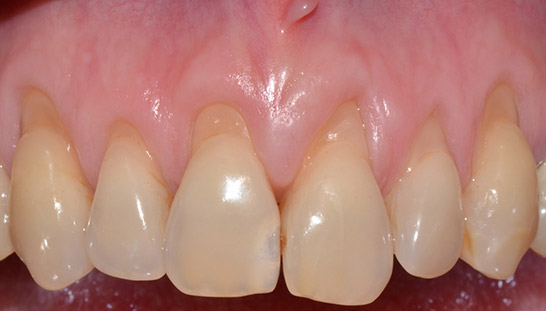
Test case: Preoperative situation.
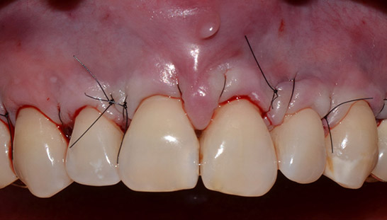
Test case: Postoperative situation after CAF performed with a tunneling procedure on the midline papilla.
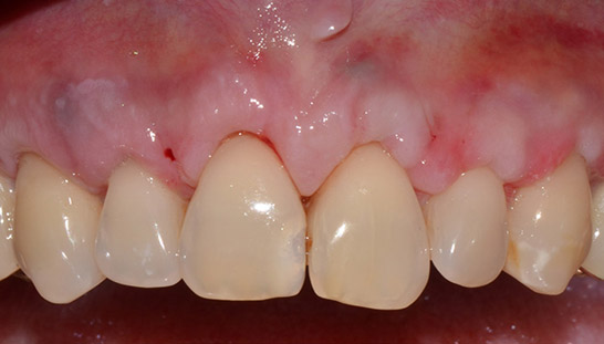
Test case: Clinical situation at seven days, immediately after suture removal.
Test case: Clinical situation at two months.
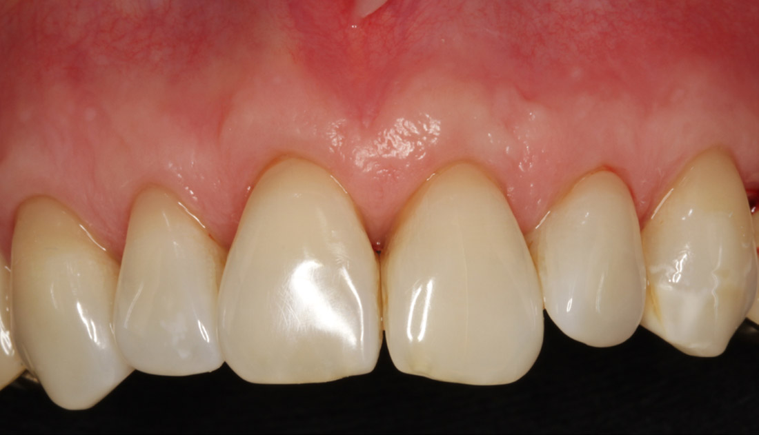
Fig. 5
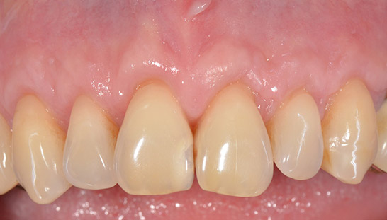
Test case: Clinical situation at one year
The flap was then secured in a coronal position, covering the cementoenamel junction of each involved tooth by suturing the papillae with synthetic monofilament 5-0 sutures (Monomyd, Butterfly Italia, Cavenago di Brianza, Italy; POLINYL, Sweden & Martina, Due Carrare, Italy; Cytoplast, Osteogenics Biomedical, Lubbock, Texas, U.S.). In the postoperative period, ketoprofen (OKi, Dompé, Milan, Italy) according to the patient’s need was prescribed for pain control. Patients were instructed to abstain from consuming hot food and beverages for two days and from chewing hard food in the area of intervention until suture removal. Equally, no flossing or brushing around the treated teeth was allowed and a 0.12% chlorhexidine spray (CURASEPT ADS Spray, Curaden, Saronno, Italy) was prescribed for local application t.i.d. after meals. After suture removal, proper oral hygiene measures were re-established, starting with brushing with an ultrasoft postoperative toothbrush. Furthermore, examinations were scheduled for 2, 4, 8 and 12 months, measuring again all preoperative clinical parameters at the 12-month control (Figs. 1–5). REC reduction (RECred) and the CRC rate for the test and control groups were calculated for all teeth involved in the treatment and for the central incisors adjacent to the midline papilla. Differences in terms of RECred and the CRC rate between the test and control groups were determined according to statistical analysis for all of the RECs by means of the Student’s t-test for independent samples and the chi-squared test, respectively, and limited to those at the central incisors by the Mann–Whitney U test and Fisher exact test, respectively. A p-value of < 0.05 was considered statistically significant.
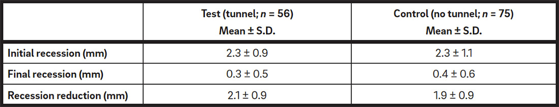
Recession reduction: Comparison between the test and control groups.

Complete root coverage: Comparison between the test and control groups.
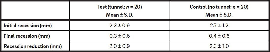
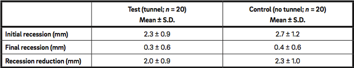
Recession reduction of central incisors: Comparison between the test and control groups.
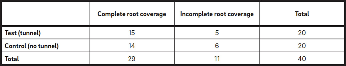
Complete root coverage of central incisors: Comparison between the test and control groups.
Results
Fifty-seven Miller Class I RECs were treated in the test group and 76 in the control group. One REC exhibiting less than 2 mm of residual keratinized tissue in each group received a connective tissue graft or a graft substitute and was not considered in the study. Therefore, 56 (mean initial REC = 2.3 ± 0.9 mm) and 75 (mean initial REC = 2.3 ± 1.1 mm) RECs were analyzed for each treatment group.
The mean final REC at 12 months was 0.3 ± 0.5mm for the test group and 0.4 ± 0.6 mm for the control, with a RECred of 2.1 ± 0.9 mm (89.1% of the initial REC) and 1.9 ± 0.9 mm (84.3% of the initial REC), respectively. The Student’s t-test for unpaired data did not find a statistically significant difference in RECred between the two groups (p = 0.9692; Table 1). Forty-three out of 56 (76.8%) RECs in the test group and 53 out of 75 (70.7%) in the control group achieved CRC. The chi-squared test did not demonstrate a statistically significant difference in the CRC rate between the two groups (p = 0.4336; Table 2).
Table 3 shows the data of the RECs at the central incisors adjacent to the tunneled or not tunneled papilla. The initial mean REC at the central incisors was 2.3 ± 0.9 mm and 2.7 ± 1.2 mm, respectively, for the test and control groups. The mean final REC after 12 months for the test and control groups was 0.3 ± 0.6 mm and 0.4 ± 0.6 mm, respectively, with a RECred from the baseline of 2.0 ± 0.9 mm (87%) for the test and 2.3 ± 1.0 mm (87%) for the control groups. The Mann–Whitney U test did not show a statistically significant difference in RECred between the two groups (p= 0.27572; Table 3). Fifteen out of 20 (75%) RECs in the test group and 14 out of 20 (70%) in the control achieved CRC. The Fisher exact test did not find a statistically significant difference in the CRC rate between the two groups (p = 0.7401; Table 4).
Discussion
The results of CAF performed with a tunneling procedure underneath the maxillary midline papilla were better in terms of RECred than those of the control group, although the differences did not achieve statistical significance. They were 89.6% aligned with the outcomes of overall periodontal plastic procedures from a recent systematic review of the literature (86.27%)15Graziani F, Gennai S, Roldán S, Discepoli N, Buti J, Madianos P, Herrera D. Efficacy of periodontal plastic procedures in the treatment of multiple gingival recessions. → J Clin Periodontol. 2014 Apr;41 Suppl 15:S63–76. and with those from another publication on CAF with no releasing incisions in the same esthetic area (89.1%).16Ozcelik O, Haytac MC, Seydaoglu G. Treatment of multiple gingival recessions using a coronally advanced flap procedure combined with button application. → J Clin Periodontol. 2011 Jun;38(6):572–80. However, limited to the same esthetic area, they were slightly inferior to those of both CAF improved with an orthodontic device for a sling suture and flap securing in a more coronal position (96.2%)17Ozcelik O, Haytac MC, Seydaoglu G. Treatment of multiple gingival recessions using a coronally advanced flap procedure combined with button application. → J Clin Periodontol. 2011 Jun;38(6):572–80. and CAF alone (95.0%),18Zucchelli G, De Sanctis M. Long-term outcome following treatment of multiple Miller class I and II recession defects in esthetic areas of the mouth. → J Periodontol. 2005 Dec;76(12):2286–92. even on monolateral RECs (97.0%)19Zucchelli G, De Sanctis M. Treatment of multiple recession-type defects in patients with esthetic demands. → J Periodontol. 2000 Sep;71(9):1506–14. or in a limited number of patients and RECs (97.0%).20Zucchelli G, De Sanctis M. The coronally advanced flap for the treatment of multiple recession defects: a modified surgical approach for the upper anterior teeth. → J Int Acad Periodontol. 2007 Jul;9(3):96–103. In this study, CRC too (76.8%) was comprised in the upper level of the range of outcomes of overall periodontal plastic procedures (23.8–89.3%)21Graziani F, Gennai S, Roldán S, Discepoli N, Buti J, Madianos P, Herrera D. Efficacy of periodontal plastic procedures in the treatment of multiple gingival recessions. → J Clin Periodontol. 2014 Apr;41 Suppl 15:S63–76. and showed better results than CRC obtained with conventional CAF with no releasing incisions in the same esthetic area (61.0%)22Ozcelik O, Haytac MC, Seydaoglu G. Treatment of multiple gingival recessions using a coronally advanced flap procedure combined with button application. → J Clin Periodontol. 2011 Jun;38(6):572–80. but worse than the outcomes obtained both with improved CAF (84.6%)23Ozcelik O, Haytac MC, Seydaoglu G. Treatment of multiple gingival recessions using a coronally advanced flap procedure combined with button application. → J Clin Periodontol. 2011 Jun;38(6):572–80. and CAF alone (84.0%; 88.0%; 89.0%)24Zucchelli G, De Sanctis M. Long-term outcome following treatment of multiple Miller class I and II recession defects in esthetic areas of the mouth. → J Periodontol. 2005 Dec;76(12):2286–92.’ 25Zucchelli G, De Sanctis M. Treatment of multiple recession-type defects in patients with esthetic demands. → J Periodontol. 2000 Sep;71(9):1506–14.’ 26Zucchelli G, De Sanctis M. The coronally advanced flap for the treatment of multiple recession defects: a modified surgical approach for the upper anterior teeth. → J Int Acad Periodontol. 2007 Jul;9(3):96–103. even within the above-mentioned limits of these last two studies.
It is important to emphasize that no previous investigation has evaluated either cases of bilateral root exposures exclusively or such a large number of consecutive RECs per patient (mean of 6.55) as in the present study. In the previously mentioned clinical studies,27Zucchelli G, De Sanctis M. Treatment of multiple recession-type defects in patients with esthetic demands. → J Periodontol. 2000 Sep;71(9):1506–14.’ 28Zucchelli G, De Sanctis M. The coronally advanced flap for the treatment of multiple recession defects: a modified surgical approach for the upper anterior teeth. → J Int Acad Periodontol. 2007 Jul;9(3):96–103.’ 29Zucchelli G, De Sanctis M. Long-term outcome following treatment of multiple Miller class I and II recession defects in esthetic areas of the mouth. → J Periodontol. 2005 Dec;76(12):2286–92. the number of consecutive RECs that underwent treatment varied with a mean of between 3.3 and 4.1 per patient. Even considering only the central incisors, the results of CAF with the tunneling procedure were better in terms of RECred and CRC than those of the control group were, although such a difference did not achieve statistical significance in this case. No comparison is possible with other investigations concerning specific data on these teeth, since the key role of this method in the symmetry and esthetics of the smile has not been reported in literature prior to this study.
Conclusion
CAF performed with tunneling of the maxillary midline papilla can be considered a minimally invasive, safe and predictable surgical procedure, but failed to demonstrate significant additional benefits in terms of RECred and CRC compared with a conventional approach in this randomized clinical trial.
Competing interests
The authors declare that they have no competing interests. The study was self-funded by the authors.
References
| 1. | ↑ | Allen EP, Miller PD Jr. Coronal positioning of existing gingiva: short term results in the treatment of shallow marginal tissue recession. → J Periodontol. 1989 Jun;60(6):316–9. |
| 2. | ↑ | Nelson SW. The subpedicle connective tissue graft. A bilaminar reconstructive procedure for the coverage of denuded root surfaces. → J Periodontol. 1987 Feb;58(2):95–102. |
| 3. | ↑ | Modica F, Del Pizzo M, Roccuzzo M, Romagnoli R. Coronally advanced flap for the treatment of buccal gingival recessions with and without enamel matrix derivative. A split-mouth study. → J Periodontol. 2000 Nov;71(11):1693–8. |
| 4. | ↑ | Harris RJ. A comparative study of root coverage obtained with an acellular dermal matrix versus a connective tissue graft: results of 107 recession defects in 50 consecutively treated patients. → Int J Periodontics Restorative Dent. 2000 Jan-Feb;20(1):51–9. |
| 5. | ↑ | McGuire MK, Scheyer ET. Xenogenic collagen matrix with coronally advanced flap compared to connective tissue with coronally advanced flap for the treatment of dehiscence-type recession defects. → J Periodontol. 2010 Aug;81(8):1108–17. |
| 6, 15, 21. | ↑ | Graziani F, Gennai S, Roldán S, Discepoli N, Buti J, Madianos P, Herrera D. Efficacy of periodontal plastic procedures in the treatment of multiple gingival recessions. → J Clin Periodontol. 2014 Apr;41 Suppl 15:S63–76. |
| 7, 13, 19, 25, 27. | ↑ | Zucchelli G, De Sanctis M. Treatment of multiple recession-type defects in patients with esthetic demands. → J Periodontol. 2000 Sep;71(9):1506–14. |
| 8. | ↑ | Zucchelli G, Mele M, Mazzotti C, Marzadori M, Montebugnoli L, De Sanctis M. Coronally advanced flap with and without vertical releasing incisions for the treatment of multiple gingival recessions: a comparative controlled randomized clinical trial. → J Periodontol. 2009 Jul;80(7):1083–94. |
| 9, 20, 26, 28. | ↑ | Zucchelli G, De Sanctis M. The coronally advanced flap for the treatment of multiple recession defects: a modified surgical approach for the upper anterior teeth. → J Int Acad Periodontol. 2007 Jul;9(3):96–103. |
| 10. | ↑ | Zuhr O, Fickl S, Wachtel H, Bolz W, Hürzeler MB. Covering of gingival recessions with a modified microsurgical tunnel technique: case report. → Int J Periodontics Restorative Dent. 2007 Sep-Oct;27(5):457–63. |
| 11. | ↑ | Aroca S, Molnár B, Windisch P, Gera I, Salvi GE, Nikolidakis D, Sculean A. Treatment of multiple adjacent miller class I and II gingival recessions with a Modified Coronally Advanced Tunnel (MCAT) technique and a collagen matrix or palatal connective tissue graft: a randomized, controlled clinical trial. → J Clin Periodontol. 2013 Jul;40(7):713–20. |
| 12, 14. | ↑ | Abundo R, Corrente G. Chirurgia plastica parodontale. Trattamento estetico delle recessioni gengivali. → 1st ed. Viterbo (Italy): ACME; c2010. Chapter #6, Tecniche chirurgiche: lembo posizionato coronalmente senza incisioni di rilascio per recessioni multiple; p. 194–243. Italian. |
| 16, 17, 22, 23. | ↑ | Ozcelik O, Haytac MC, Seydaoglu G. Treatment of multiple gingival recessions using a coronally advanced flap procedure combined with button application. → J Clin Periodontol. 2011 Jun;38(6):572–80. |
| 18, 24, 29. | ↑ | Zucchelli G, De Sanctis M. Long-term outcome following treatment of multiple Miller class I and II recession defects in esthetic areas of the mouth. → J Periodontol. 2005 Dec;76(12):2286–92. |


Leave a Reply
Be the First to Comment!