Influence of fatigue on resistance and deformation of implant abutments used for provisional prosthetic restoration
September 21, 2016 / Categories: Digital Dentistry, Implant Dentistry

Agustín Panadero, Rubén

Fons Font, Antonio

Peñarrocha Oltra, David

Solá Ruíz, María Fernanda
One of the most di icult challenges for implant-supported prosthetic restorations is the management and maintenance of periimplant soft- tissue esthetics, especially in the anterior region. In order to do this e ectively, there are diverse techniques for tissue management, the most significant being fixed provisionalization, which can be immediate or delayed.
Introduction
The aim of this study was to analyze fracture resistance of provisional implant-prosthetic abutments (titanium, PEEK and methacrylate) and to determine whether previously fatiguing the abutments influenced fracture resistance.
In the field of dentistry, implant dentistry is one area that has undergone extensive development in recent years, owing to the high demand for this treatment and constant innovation and research into new materials and attachments. Implant placement has become the first treatment choice for replacing missing teeth, particularly single teeth, because of the excellent clinical results confirmed by long-term research.1Jung RE, Zembic A, Pjetursson BE, Zwahlen M, Thoma DS. Systematic review of the survival rate and the incidence of biological, technical, and aesthetic complications of single crowns on implants reported in longitudinal studies with a mean follow-up of 5 years.
→ Clin Oral Implants Res. 2012 Oct;23 Suppl 6:2–21. Nowadays, esthetics is an important factor in judging the final outcome of dental treatment. In the case of implant dentistry, various factors influence esthetics. It is not enough to place a natural-looking restoration with correct proportions and adequate color, for a successful outcome will also depend on management of the periimplant soft tissue.2Bruno V, Badino M, Sacco R, Catapano S. The use of a prosthetic template to maintain the papilla in the esthetic zone for immediate implant placement by means of a radiographic procedure.
→ J Prosthet Dent. 2012 Dec;108(6):394–7.This is not always straightforward, as the soft tissue is governed by multiple factors: the periodontal biotype, alveolar bone crest level, angle of implant insertion, depth of implant platform and level of the first point of bone to implant contact.3Nisapakultorn K, Suphanantachat S, Silko-Sessak O, Rattanamongkolgul S. Factors a ecting soft tissue level around anterior maxillary single tooth implants. → Clin Oral Implants Res. 2010 Jun;21(6):662–70. Given this scenario, achieving optimal esthetic results is a complicated process. A diverse range of techniques are available for soft-tissue management. From the prosthodontic perspective, provisional prostheses are useful to help model the surrounding tissue and create a harmonious profile before placing the definitive restoration.4Manicone PF, Ra aelli L, Ghassemian M, D’Addona A. Soft and hard tissue management in implant therapy—Part II: prosthetic concepts.
→ Int J Biomater. 2012;2012:356817. Epub 2012 Jul 3.Furthermore, provisional restorations help improve communication with the patient, as they o er the opportunity to view future outcomes.5Bruno V, Badino M, Sacco R, Catapano S. The use of a prosthetic template to maintain the papilla in the esthetic zone for immediate implant placement by means of a radiographic procedure.
→ J Prosthet Dent. 2012 Dec;108(6):394–7.
For all these reasons, dental professionals need to be aware of the di erent materials available on the market, as well as their physical and chemical properties, for the correct fabrication of both provisional and definitive prostheses that will achieve optimal esthetics and good peri implant health.6Nisapakultorn K, Suphanantachat S, Silko-Sessak O, Rattanamongkolgul S. Factors a ecting soft tissue level around anterior maxillary single-tooth implants. → Clin Oral Implants Res. 2010 Jun;21(6):662–70.7Manicone PF, Ra aelli L, Ghassemian M, D’Addona A. Soft and hard tissue management in implant therapy—Part II: prosthetic concepts.
→ Int J Biomater. 2012;2012:356817. Epub 2012 Jul 3.8Agustín-Panadero R, Serra-Pastor B, Roig-Vanaclocha A, Román-Rodriguez JL, Fons-Font A. Mechanical behavior of provisional implant prosthetic abutments. → Med Oral Patol Oral Cir Bucal. 2015 Jan;20(1):e94–102.9Misch CE, Wang HL, Misch CM, Sharawy M, Lemons J, Judy KW. Rationale for the application of immediate load in implant dentistry: Part II.
→ Implant Dent. 2004 Dec;13(4):310–21.10Odin G, Misch CE, Binderman I, Scortecci G. Fixed rehabilitation of severely atrophic jaws using immediately loaded basal disk implants after in situ bone activation.
→ J Oral Implantol. 2012 Oct;38(5):611–6.11Chung DM, Oh TJ, Lee J, Misch CE, Wang HL. Factors a ecting late implant bone loss:
a retrospective analysis.
→ Int J Oral Maxillofac Implants. 2007 Jan-Feb;22(1):117–26.12Misch CE. Consideration of biomechanical stress in treatment with dental implants.
→ Dent Today.2006 May;25(5):80, 82, 84–5; quiz 85.13Van der Weijden F, Dell’Acqua F, Slot DE. Alveolar bone dimensional changes of post-extraction sockets in humans: a systematic review.
→ J Clin Periodontol. 2009 Dec;36(12):1048–58.14Schropp L, Wenzel A, Kostopoulos L, Karring T. Bone healing and soft tissue contour changes following single-tooth extraction:
a clinical and radiographic 12-month prospective study.
→ Int J Periodontics Restorative Dent. 2003 Aug;23(4):313–23. Provisional restoration can be useful as a diagnostic tool too, as it allows the dentist to assess the final outcome in advance and provides an opportunity to obtain the patient’s feedback and opinion. Its main function is to guide and shape the soft tissue during healing and maturation, allowing the tissue to develop more quickly and suggest the definitive gingival shape.15Araújo MG, Lindhe J. Dimensional ridge alterations following tooth extraction. An experimental study in the dog.
→ J Clin Periodontol. 2005 Feb;32(2):212–8.16Von Wowern N, Gotfredsen K. Implant-sup- ported overdentures, a prevention of bone loss in edentulous mandibles? A 5-year follow-up study.
→ Clin Oral Implants Res. 2001 Feb;12(1):19–25.17Herrera Briones FJ, Romero Oild MN, Vallecillo Capilla M. Puesta al día sobre implantes de carga inmediata. Revisión bibliográfica.
→ Med Oral Patol Oral Cir Bucal. 2004 Feb;9(1):74–81. Spanish.18Maló P, Rangert B, Nobre M. “All-on-Four” immediate-function concept with Brånemark System implants for completely edentulous mandibles: a retrospective clinical study.
→ Clin Implant Dent Relat Res. 2003 Mar;5 Suppl 1:2–9.19Maló P, Friberg B, Polizzi G, Gualini F, Vighagen T, Rangert B. Immediate and early function of Brånemark System implants placed in the esthetic zone: a 1-year prospective clinical multicenter study.
→ Clin Implant Dent Relat Res. 2003 Mar;5 Suppl 1:37–46.20Cooper LF, Rahman A, Moriarty J, Cha ee N, Sacco D. Immediate mandibular rehabilitation with endosseous implants: simultaneous extraction, implant placement, and loading. → Int J Oral Maxillofac Implants. 2002 Jul-Aug;17(4):517–25.21Santosa RE. Provisional restoration options in implant dentistry.
→ Aust Dent J. 2007 Sep;52(3):234–42; quiz 254.22Canullo L, Peñarrocha-Oltra D, Soldini C, Mazzocco F, Peñarrocha M, Covani U. Microbiological assessment of the implant-abutment interface in di erent connections: cross-sectional study after 5 years of functional loading.
→ Clin Oral Implants Res. 2015 Apr;26(4):426–34.23Uribe R, Peñarrocha M, Balaguer J, Fulgueiras N. Carga inmediata en implantología oral. Situación actual [Immediate loading in oral implants. Present situation].
→ Med Oral Patol Oral Cir Bucal. 2005 Jul;10 Suppl 2:E143–53. Spanish.24Binon PP. Implants and components: entering the new millennium.
→ Int J Oral Maxillofac Implants. 2000 Jan-Feb;15(1):76–94.25Peñarrocha-Diago MA, Flichy-Fernandez AJ, Alonso-González R, Peñarrocha-Oltra D, Balaguer-Martínez J, Peñarrocha-Diago M. Influence of implant neck design and implant-abutment connection type on peri-implant health. Radiological study.
→ Clin Oral Implants Res. 2013 Nov;24(11):1192–200.26Canullo L, Pace F, Coelho P, Sciubba E, Vozza I. The influence of platform switching on the biomechanical aspects of the implant-abutment system. A three dimensional finite element study.
→ Med Oral Patol Oral Cir Bucal. 2011 Sep;16(6):e852–6.27Sailer I, Mühlemann S, Zwahlen M, Hämmerle CH, Schneider D. Cemented and screw-re- tained implant reconstructions: a systematic review of the survival and complication rates. → Clin Oral Implants Res. 2012 Oct;23 Suppl 6:163–201.28Priest G. Esthetic potential of single-implant provisional restorations: selection criteria of available alternatives.
→ J Esthet Restor Dent. 2006 Nov-Dec;18(6):326–38; discussion 339.29Lewis MB, Klineberg I. Prosthodontic considerations designed to optimize outcomes for single-tooth implants. A review of the literature.
→ Aust Dent J. 2011 Jun;56(2):181–92.30Shemtov-Yona K, Rittel D, Levin L, Matchetei EE. The e ect of oral-like environment on dental implants’ fatigue performance.
→ Clin Oral Implants Res. 2014 Feb;25(2):e166–70.31Steinebrunner L, Wolfart S, Ludwig K, Kern M. Implant abutment interface design a ects fatigue and fracture strength of implants.
→ Clin Oral Implants Res. 2008 Dec;19(12):1276–84.32Peñarrocha-Oltra D, Covani U, Peñarrocha M, Peñarrocha-Diago M. Immediate versus conventional loading with fixed full-arch prostheses in mandibles with failing dentition: a prospective controlled study.
→ Int J Oral Maxillofac Implants. 2015 Mar-Apr;30(2):427–34.
This study was designed with the following objectives:
– to analyze the deformation and fracture resistance of implant-supported provisional abutments made of di erent materials (titanium, PEEK and methacrylate)
– to determine whether fatiguing prior to static load testing influenced fracture resistance and deformation of the abutments.
Materials and methods
Materials
Forty Kohno internal hex connection implants (Sweden & Martina, Due Carrare, Italy) were used (4.25 mm in diameter and 11.5 mm in length). Forty abutments were screwed on to the implants, 30 of which were provisional and ten definitive (n = 40). The abutments were divided into four groups (Table 1): castable methacrylate provisional (CMP) abutments with a titanium base; PEEK (polyether ether ketone) provisional (PP) abutments with a machined titanium base; Grade III titanium provisional (TP) abutments; and Grade IV titanium definitive (TD) abutments.
Forty specimens were fabricated, each consisting of an implant set in a 5 cm diameter nylon cylinder with epoxy resin (Exakto-Form, bredent, Senden, Germany). In order to simulate implant position conditions in the alveolar ridge in the premaxilla, specimens were placed in the cylinder at an angle of 30° to the direction of the load. The abutments were screwed on to the implant– cylinder complex using a dynamometric torque wrench, applying a torque of 30 N, as recommended by the manufacturer.
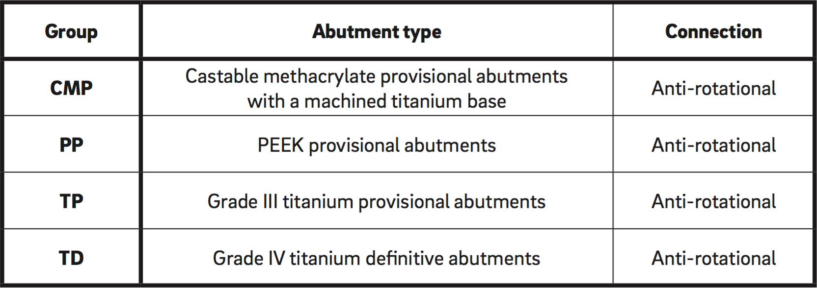
Table. 1
Specimen distribution by abutment type
Fig. 1
Cyclic loading of implant- supported abutments.
Fig. 2
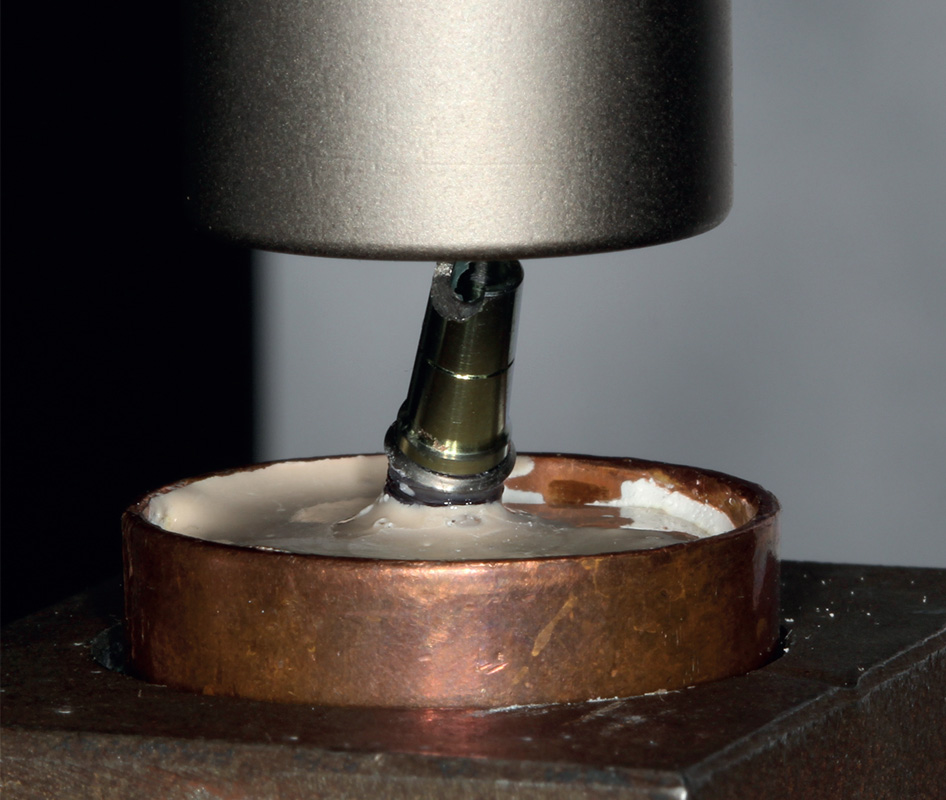
Static load testing of implant-supported abutments.
Method
Before specimens underwent static load testing, they were subjected to dynamic loading. This fatiguing process was performed using a chewing simulator
(CS-4, SD Mechatronik. Rosenheim, Germany; Fig. 1). Loading was applied to the upper part of the abutment (angled at 30°) with an impact force of 80 N and a frequency of 2 Hz. Each specimen was subjected to 60,000 cycles at an application speed of 40 mm/s. They then underwent thermocycling (Thermocycler 2000, Heto-Holten A/S, Allerod, Denmark) for 6000 cycles with temperature changes between 5°C and 55°C every 30 seconds.
Static compression load testing was used to evaluate the abutments’ fracture resistance. The testing was performed using a static load testing machine (AG-X plus, Shimadzu, Kyoto, Japan). A load cell of 5,000 N was used at a crosshead speed of 0.5 mm/min
(Fig. 2).
Statistical analysis consisted of preliminary descriptive analysis of the force (fracture resistance) and deformation variables (mean, standard deviation, range and median). Comparisons were made adopting a nonparametric approach. Significance was set at 5% (p = 0.05).
-

- Fig.1
-
- Fig.2
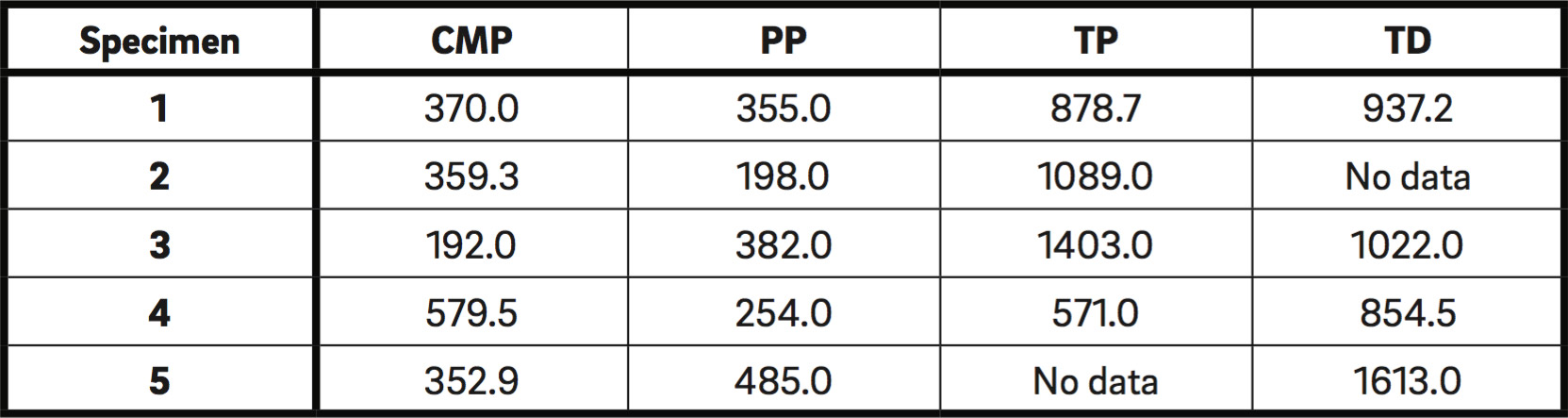
Table. 2
Fracture resistance (N) for implant-prosthetic abutments not subjected to fatiguing.
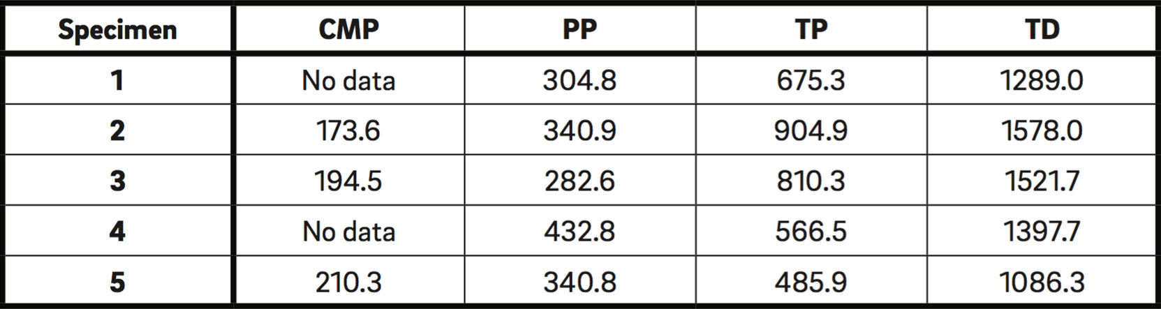
Table. 3
Fracture resistance (N) for implant-prosthetic abutments subjected to fatiguing.
Table. 4
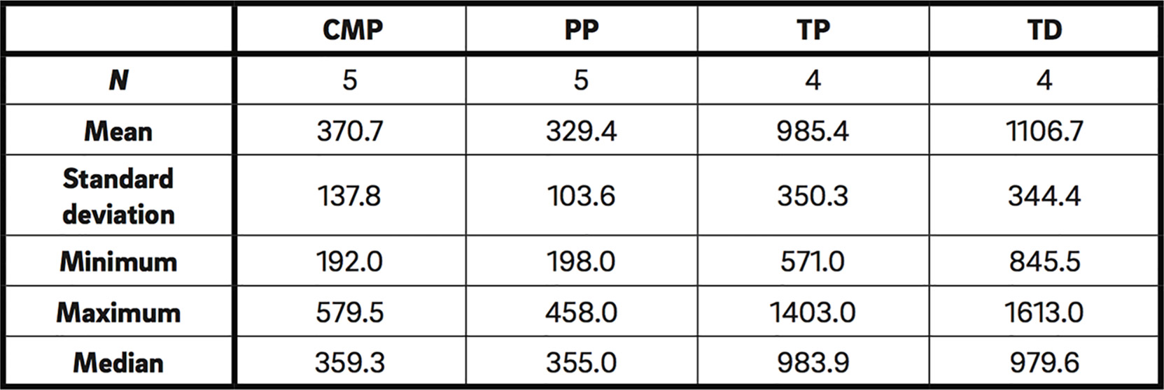
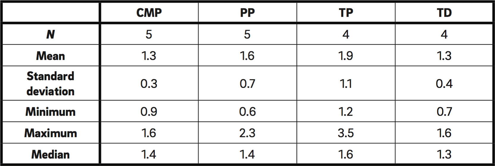
Descriptive data by group for abutments not subjected to fatiguing (N).
Table. 5
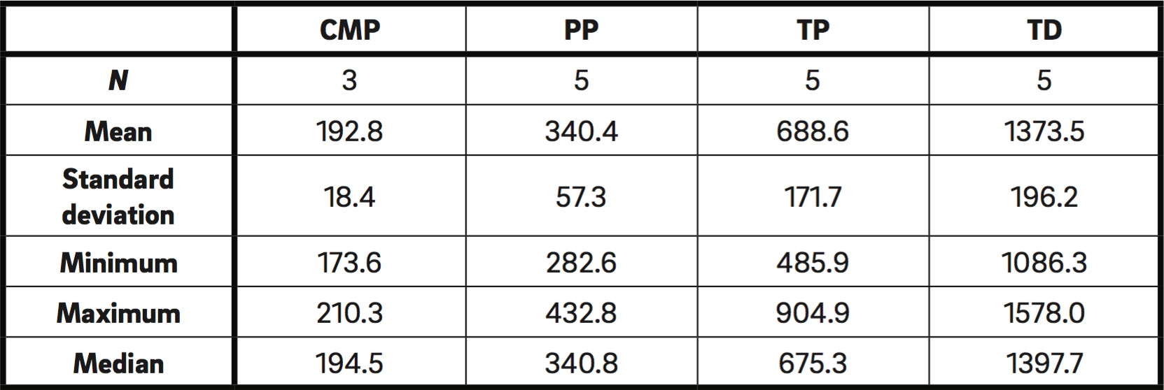
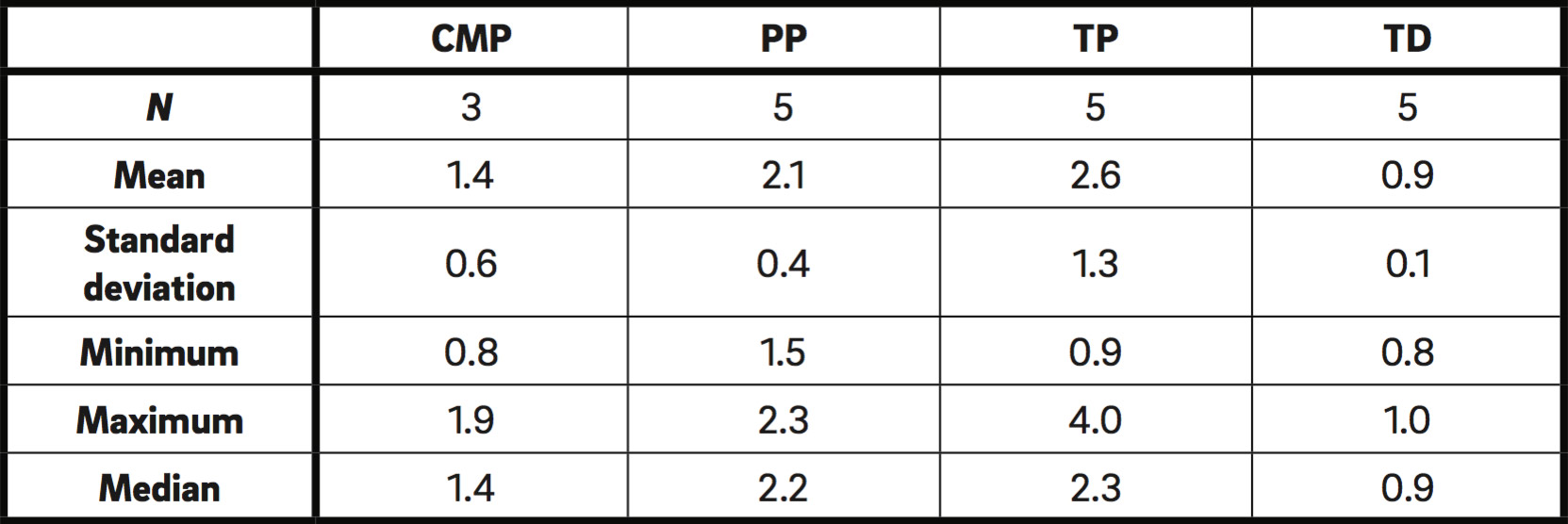
Descriptive data by group for abutments subjected to fatiguing (N).
Table. 6
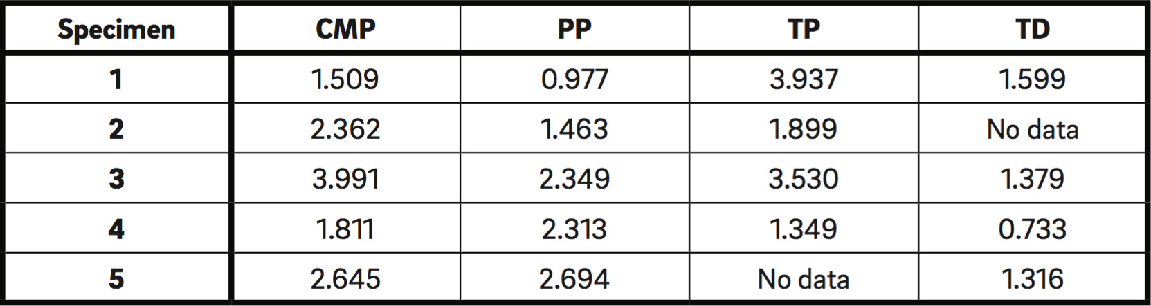
Deformation data (mm).
Table. 7
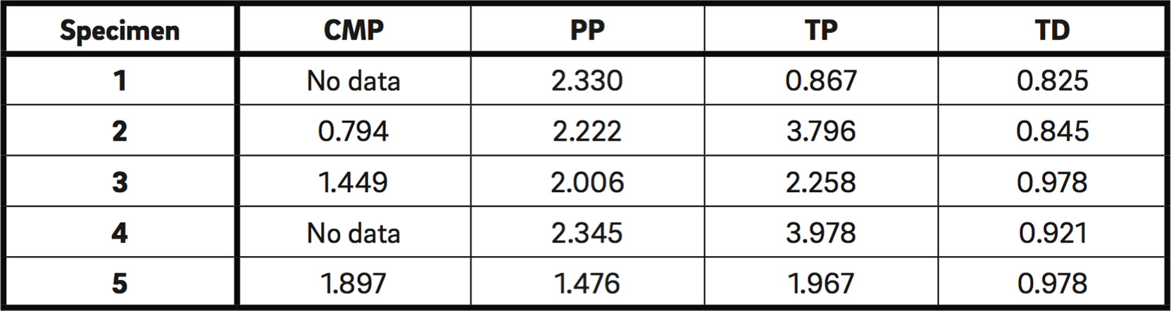
Deformation data (mm).
Table. 8
Descriptive deformation data by group for abutments not subjected to fatiguing (mm).
Table. 9
Descriptive deformation data by group for abutments subjected to fatiguing (mm).
Results
The results obtained registered the force in Newtons (N) required to produce the fracture of each specimen (Tables 2 & 3). Fracture of the prosthesis was understood as the first mechanical failure that the specimen underwent, whether this was the maximum load that produced a clearly observed fracture or the maximum load before the test machine registered a decrease in load even if the fracture was not visibly obvious. Fracture resistance values for two specimens (not subjected to fatiguing) were discarded owing to failure to fulfill the study procedure. The same also occurred with two specimens subjected to fatiguing.
Table 4 shows the descriptive data by group for fracture resistance in specimens not subjected to fatiguing. The group that presented the highest resistance to fracture was the TD group and the group that showed the least resistance was the PP group, with mean values of 1,106.7 N and 329.4 N, respectively. The groups that presented the lowest resistance to fracture were CMP and PP, obtaining values of between 300 N and 400 N. Fracture resistance levels were heterogeneous, as the Kruskal–Wallis test confirmed that there was no homogeneity in the distribution of resistance across the four groups (p = 0.006).
When the Mann–Whitney test was applied to identify di erences between pairs of groups, CMP showed lower resistance than TP (p = 0.032) and TD (p = 0.016), with the di erences being statistically significant. PP restorations obtained lower resistance than the TP (p = 0.016) and TD groups (p = 0.016), with the di erences being statistically significant. The performance of the TP group was similar to that of the TD group (p = 0.886). CMP and PP were also homogenous, but with a significantly lower resistance than the other two groups (p = 1.000).
In a comparison of the statistical data for fracture resistance of restorations subjected to fatiguing (Table 5), the group that showed the highest resistance was the TD group (1,373.5 N). The groups with the lowest resistance were PP and CMP; both groups obtained values of between 200 N and 350 N. Fracture resistance levels were heterogeneous, and the Kruskal– Wallis test confirmed that there was no heterogeneity in the distribution of resistance across the four groups (p < 0.001).
When resistance distribution was compared between pairs of groups of fatigued specimens,
statistically significant di erences were identified for all comparisons. Unlike the groups not subjected to fatiguing, no group of fatigued specimens presented a homogenous distribution of resistance when paired comparisons were made.
CMP restorations showed lower fracture resistance than the rest of the groups, with the differences being statistically significant (PP: p = 0.036; TP: p = 0.036; TD: p = 0.036). The PP group also showed lower resistance than TP (p = 0.008) and TD specimens (p = 0.008), with the di erences being statistically significant.
In making a comparative analysis between the specimens subjected to fatiguing and those that were not fatigued, a slight decrease in fracture resistance was observed among all of the provisional restorations subjected to fatiguing (CMP, PP and TP). However, the TD group showed stable performance despite cyclic loading, such that its performance was not a ected by fatiguing. The Mann–Whitney test was applied to evaluate di erences between fatigued and nonfatigued specimens and a p-value of 0.401 was obtained, indicating that resistance to fracture was similar between the two groups (fatigued/ nonfatigued). Likewise, within the individual groups, no significant di erences were found between fatigued or nonfatigued subgroups (CMP: p = 0.143; PP: p 1.000; TP: p = 0.190; TD: p 0.286), although the CMP and TP groups did show a certain tendency toward di erence, but this did not reach statistical significance.
In data analysis of the deformation that the restorations su ered up to the point of fracture or mechanical failure (in mm; Tables 6 & 7), deformation values for specimen 2 in the TD group and specimen 5 in the TP group (not fatigued) were discarded from analysis owing to various technical failures in the study procedure. The same occurred with two specimens (1 and 4) in the CMP group (subjected to fatiguing).In deformation data analysis of groups not subjected to fatiguing (Table 8), it was found that the group that presented the highest deformation values was the TP group. The group with the least deformation was the TD group. The PP and CMP groups showed similar median values, but a dispersed range of values. The Kruskal–Wallis test showed that there were no overall significant di erences (p = 0.187). The Mann–Whitney test found that the only significant difference occurred between the CMP and TD groups, the TD group showing the lowest deformation values (CMP–PP: p = 0.421; CMP–TP: p = 1.000; PP–TP: p = 0.556; CMP–TD: p = 0.032; PP–TD: p = 0.190; TP–TD: p = 0.114).
As for deformation data analysis of specimens subjected to fatiguing (Table 9), the TD and CMP groups underwent the least deformation. The PP and TP groups showed similar median values, but the range of values was more widely dispersed in the TP group. The group that underwent the greatest deformation was the TP group. When homogeneity was analyzed between groups, the Kruskal–Wallis test found a p-value of 0.022, indicating homogeneity between the groups. The Mann–Whitney test for paired groups only identified statistically significant di erences between the TD and PP groups, with the TD group obtaining lower deformation values (CMP–PP: p = 0.071; CMP–TP: p = 0.143; PP–TP: p = 0.841; CMP–TD: p = 0.571; PP–TD: p = 0.008; TP–TD: p = 0.056).
In order to determine whether fatigue influenced deformation, specimens subjected to fatiguing were compared with those not subjected to fatiguing, but no significant di erences were found (CMP: p = 0.143; PP: p = 1.000; TP: p = 0.905; TD: p = 0.286).
Discussion
Nowadays, many patients regard dental esthetics as one of the principal requirements of dental treatment. In the case of implant dentistry, a range of factors influence esthetic outcomes, including color, contour, the natural appearance of the definitive prosthesis, and most importantly, the topography and appearance of the periimplant soft tissue.33Bruno V, Badino M, Sacco R, Catapano S. The use of a prosthetic template to maintain the papilla in the esthetic zone for immediate implant placement by means of a radiographic procedure.
→ J Prosthet Dent.2012 Dec;108(6):394–7. Soft-tissue management is not straightforward, as multiple factors a ect the final outcome, in which the provisional prosthesis plays a key role.34Bruno V, Badino M, Sacco R, Catapano S. The use of a prosthetic template to maintain the papilla in the esthetic zone for immediate implant placement by means of a radiographic procedure.
→ J Prosthet Dent.2012 Dec;108(6):394–7.35Manicone PF, Ra aelli L, Ghassemian M, D’Addona A. Soft and hard tissue management in implant therapy—Part II: prosthetic concepts.
→ Int J Biomater. 2012;2012:356817. Epub 2012 Jul 3. Given the importance of provisionalization as a part of dental implant treatment, the present study set out to evaluate the resistance to fracture of implant-supported provisional prostheses of di erent materials (titanium, PEEK resin and methacrylate) subjected to fatiguing. While definitive prostheses have been extensively studied, little research has investigated fracture resistance and the influence of fatigue on provisional abutments in vitro.
The present study protocol was designed to fulfill the test geometry specified in ISO 14801:2007 for testing single-post endosseous dental implants, in that the implant made a 30° angle with the test machine’s load cell.36Martínez-Rus F, Ferreiroa A, Özcan M, Bartolomé JF, Pradíes G. Fracture resistance of crowns cemented on titanium and zirconia implant abutments: a comparison of monolithic versus manually veneered all-ceramic systems.
→ Int J Oral Maxillofac Implants. 2012 Nov-Dec;27(6):1448–55.37Nothdurf FP, Doppler KE, Erdelt KJ, Knauber AW, Pospiech PR. Fracture behavior of straight or angulated zirconia implant abutments supporting anterior single crowns. → Clin Oral Investig. 2011 Apr;15(2):157–6338Nothdurf FP, Doppler KE, Erdelt KJ, Knauber AW, Pospiech PR. Influence of artificial aging on the load-bearing capability of straight or angulated zirconia abutments in implant/ tooth-supported fixed partial dentures.
→ Int J Oral Maxillofac Implants. 2010 Sep;25(5):991–8.39Rack T, Zabler S, Rack A, Riesemeier H, Nelson K. An in vitro pilot study of abutment stability during loading in new and fatigue-loaded conical dental implants using synchrotron- base radiography.
→ Int J Oral Maxillofac Implants. 2013 Jan-Feb;28(1):44–50.40Sannino G, Barlattani A. Mechanical evaluation of an implant-abutment self-locking connection: finite element analysis and experimental test.
→ Int J Oral Maxillofac Implants. 2013 Jan-Feb;28(1):e17–26.41Truninger TC, Stawarczyk B, Leutert CR, Sailer TR, Hämmerle CH, Sailer I. Bending moments of zirconia and titanium abutments with internal and external implant-abutment connections after aging and chewing simulation.
→ Clin Oral Implants Res. 2012 Jan;23(1):12–8.42Stimmelmayr M, Sagerer S, Erdelt K, Beuer F. In vitro fatigue and fracture strength testing of one piece zirconia implant abutment and zirconia implant abutments connected to titanium cores.
→ Int J Oral Maxillofac Implants. 2013 Mar-Apr;28(2):488–93.43Simsiriwong J, Shrestha R, Shamsaei N, Lugo M, Moser RD. E ects of microstructural inclusions on fatigue life of polyether ether ketone (PEEK).
→ J Mech Behav Biomed Mater. 2015 Nov;51:388–97.44Ferrario VF, Sforza C, Serrao G, Dellavia C, Tartaglia GM. Single tooth bite forces in healthy young adults.
→ J Oral Rehabil. 2004 Jan;31(1):18–22.45International Organization for Standardizati- on. International standard ISO 14801:2007: dentistry—implants—dynamic fatigue test for endosseous dental implants.
→ Geneva: International Organization for Standardization, 2007. 9 p. This geometry has been used in most other studies of similar characteristics to the present one.46Sannino G, Barlattani A. Mechanical evaluation of an implant-abutment self-locking connection: finite element analysis and experimental test.
→ Int J Oral Maxillofac Implants. 2013 Jan-Feb;28(1):e17–26.47Truninger TC, Stawarczyk B, Leutert CR, Sailer TR, Hämmerle CH, Sailer I. Bending moments of zirconia and titanium abutments with internal and external implant-abutment connections after aging and chewing simulation.
→ Clin Oral Implants Res. 2012 Jan;23(1):12–8.48Stimmelmayr M, Sagerer S, Erdelt K, Beuer F. In vitro fatigue and fracture strength testing of one piece zirconia implant abutment and zirconia implant abutments connected to titanium cores.
→ Int J Oral Maxillofac Implants. 2013 Mar-Apr;28(2):488–93.49Simsiriwong J, Shrestha R, Shamsaei N, Lugo M, Moser RD. E ects of microstructural inclusions on fatigue life of polyether ether ketone (PEEK).
→ J Mech Behav Biomed Mater. 2015 Nov;51:388–97. The material used to set the implant in the cylinder—epoxy resin—was chosen for its elastic modulus > 3 GPa, also required by ISO 14801:2007, and because this material has been used in similar studies too.50Martínez-Rus F, Ferreiroa A, Özcan M, Bartolomé JF, Pradíes G. Fracture resistance of crowns cemented on titanium and zirconia implant abutments: a comparison of monolithic versus manually veneered all-ceramic systems.
→ Int J Oral Maxillofac Implants. 2012 Nov-Dec;27(6):1448–55.51Nothdurf FP, Doppler KE, Erdelt KJ, Knauber AW, Pospiech PR. Fracture behavior of straight or angulated zirconia implant abutments supporting anterior single crowns. → Clin Oral Investig. 2011 Apr;15(2):157–63.52Nothdurf FP, Doppler KE, Erdelt KJ, Knauber AW, Pospiech PR. Influence of artificial aging on the load-bearing capability of straight or angulated zirconia abutments in implant/ tooth-supported fixed partial dentures.
→ Int J Oral Maxillofac Implants. 2010 Sep;25(5):991–8.53Rack T, Zabler S, Rack A, Riesemeier H, Nelson K. An in vitro pilot study of abutment stability during loading in new and fatigue-loaded conical dental implants using synchrotron- base radiography.
→ Int J Oral Maxillofac Implants. 2013 Jan-Feb;28(1):44–50.54Sannino G, Barlattani A. Mechanical evaluation of an implant-abutment self-locking connection: finite element analysis and experimental test.
→ Int J Oral Maxillofac Implants. 2013 Jan-Feb;28(1):e17–26.55Truninger TC, Stawarczyk B, Leutert CR, Sailer TR, Hämmerle CH, Sailer I. Bending moments of zirconia and titanium abutments with internal and external implant-abutment connections after aging and chewing simulation.
→ Clin Oral Implants Res. 2012 Jan;23(1):12–8.56Stimmelmayr M, Sagerer S, Erdelt K, Beuer F. In vitro fatigue and fracture strength testing of one piece zirconia implant abutment and zirconia implant abutments connected to titanium cores.
→ Int J Oral Maxillofac Implants. 2013 Mar-Apr;28(2):488–93.57Simsiriwong J, Shrestha R, Shamsaei N, Lugo M, Moser RD. E ects of microstructural inclusions on fatigue life of polyether ether ketone (PEEK).
→ J Mech Behav Biomed Mater. 2015 Nov;51:388–97. All of the abutments were tested without placing restorations on them, as was the case in Truninger et al., in which the abutments were subjected to load testing without bearing restorations.58Truninger TC, Stawarczyk B, Leutert CR, Sailer TR, Hämmerle CH, Sailer I. Bending moments of zirconia and titanium abutments with internal and external implant-abutment connections after aging and chewing simulation.
→ Clin Oral Implants Res. 2012 Jan;23(1):12–8.Likewise, Rack et al. tested abutments without placing restorations on them, but soldered a steel sphere of 10 mm in diameter to the coronal part of the abutment so that the force applied would be evenly distributed throughout the abutment structure.59Rack T, Zabler S, Rack A, Riesemeier H, Nelson K. An in vitro pilot study of abutment stability during loading in new and fatigue-loaded conical dental implants using synchrotron- base radiography.
→ Int J Oral Maxillofac Implants. 2013 Jan-Feb;28(1):44–50.
The choice of test design was based on Agustín-Panadero et al., who studied provision-al abutments subjected to static loading.60Agustín-Panadero R, Serra-Pastor B, Roig-Vanaclocha A, Román-Rodriguez JL, Fons-Font A. Mechanical behavior of provisional implant prosthetic abutments. → Med Oral Patol Oral Cir Bucal. 2015 Jan;20(1):e94–102. Various authors have proposed similar variables to the present test design in terms of specimen design and distribution, as well as crosshead speed and movement.61Martínez-Rus F, Ferreiroa A, Özcan M, Bartolomé JF, Pradíes G. Fracture resistance of crowns cemented on titanium and zirconia implant abutments: a comparison of monolithic versus manually veneered all-ceramic systems.
→ Int J Oral Maxillofac Implants. 2012 Nov-Dec;27(6):1448–55.62Nothdurf FP, Doppler KE, Erdelt KJ, Knauber AW, Pospiech PR. Fracture behavior of straight or angulated zirconia implant abutments supporting anterior single crowns. → Clin Oral Investig. 2011 Apr;15(2):157–63.63Nothdurf FP, Doppler KE, Erdelt KJ, Knauber AW, Pospiech PR. Influence of artificial aging on the load-bearing capability of straight or angulated zirconia abutments in implant/ tooth-supported fixed partial dentures.
→ Int J Oral Maxillofac Implants. 2010 Sep;25(5):991–8.64Rack T, Zabler S, Rack A, Riesemeier H, Nelson K. An in vitro pilot study of abutment stability during loading in new and fatigue-loaded conical dental implants using synchrotron- base radiography.
→ Int J Oral Maxillofac Implants. 2013 Jan-Feb;28(1):44–50.65Sannino G, Barlattani A. Mechanical evaluation of an implant-abutment self-locking connection: finite element analysis and experimental test.
→ Int J Oral Maxillofac Implants. 2013 Jan-Feb;28(1):e17–26.66Truninger TC, Stawarczyk B, Leutert CR, Sailer TR, Hämmerle CH, Sailer I. Bending moments of zirconia and titanium abutments with internal and external implant-abutment connections after aging and chewing simulation.
→ Clin Oral Implants Res. 2012 Jan;23(1):12–8.The crosshead speed in compression testing in this study was 0.5 mm/min, a speed established from the literature review conducted in preparation for the study to ensure use of the same speed used in the majority of other similar studies (standardization being important when it comes to comparison of studies).67Martínez-Rus F, Ferreiroa A, Özcan M, Bartolomé JF, Pradíes G. Fracture resistance of crowns cemented on titanium and zirconia implant abutments: a comparison of monolithic versus manually veneered all-ceramic systems.
→ Int J Oral Maxillofac Implants. 2012 Nov-Dec;27(6):1448–55.68Nothdurf FP, Doppler KE, Erdelt KJ, Knauber AW, Pospiech PR. Fracture behavior of straight or angulated zirconia implant abutments supporting anterior single crowns. → Clin Oral Investig. 2011 Apr;15(2):157–63.69Nothdurf FP, Doppler KE, Erdelt KJ, Knauber AW, Pospiech PR. Influence of artificial aging on the load-bearing capability of straight or angulated zirconia abutments in implant/ tooth-supported fixed partial dentures.
→ Int J Oral Maxillofac Implants. 2010 Sep;25(5):991–8.70Rack T, Zabler S, Rack A, Riesemeier H, Nelson K. An in vitro pilot study of abutment stability during loading in new and fatigue-loaded conical dental implants using synchrotronbase radiography.
→ Int J Oral Maxillofac Implants. 2013 Jan-Feb;28(1):44–50.71Sannino G, Barlattani A. Mechanical evaluation of an implant-abutment self-locking connection: finite element analysis and experimental test.
→ Int J Oral Maxillofac Implants. 2013 Jan-Feb;28(1):e17–26.72Truninger TC, Stawarczyk B, Leutert CR, Sailer TR, Hämmerle CH, Sailer I. Bending moments of zirconia and titanium abutments with internal and external implant-abutment connections after aging and chewing simulation.
→ Clin Oral Implants Res. 2012 Jan;23(1):12–8.73Stimmelmayr M, Sagerer S, Erdelt K, Beuer F. In vitro fatigue and fracture strength testing of one piece zirconia implant abutment and zirconia implant abutments connected to titanium cores.
→ Int J Oral Maxillofac Implants. 2013 Mar-Apr;28(2):488–93.However, pure compression studies do appear to be adequate for researching the fracture resistance of implant-prosthetic structures. The ideal procedure in a study of these characteristics is to subject specimens to dynamic loading– artificial aging of the specimens–before performing the static load testing. For this reason, the present study divided the specimens (n = 40) into two subgroups and subjected half to a prior fatiguing process to simulate the aging of the abutments. Like compression testing, the fatiguing process must meet criteria established in ISO 14801:2007.74International Organization for Standardizati- on. International standard ISO 14801:2007: dentistry—implants—dynamic fatigue test for endosseous dental implants.
→ Geneva: International Organization for Standardization, 2007. 9 p.
The literature contains several studies that have subjected specimens to aging prior to testing.75Shemtov-Yona K, Rittel D, Levin L, Matchetei EE. The e ect of oral-like environment on dental implants’ fatigue performance.
→ Clin Oral Implants Res. 2014 Feb;25(2):e166–70.76Steinebrunner L, Wolfart S, Ludwig K, Kern M. Implant abutment interface design a ects fatigue and fracture strength of implants.
→ Clin Oral Implants Res. 2008 Dec;19(12):1276–84. 77Martínez-Rus F, Ferreiroa A, Özcan M, Bartolomé JF, Pradíes G. Fracture resistance of crowns cemented on titanium and zirconia implant abutments: a comparison of monolithic versus manually veneered all-ceramic systems.
→ Int J Oral Maxillofac Implants. 2012 Nov-Dec;27(6):1448–55. 78Sannino G, Barlattani A. Mechanical evaluation of an implant-abutment self-locking connection: finite element analysis and experimental test.
→ Int J Oral Maxillofac Implants. 2013 Jan-Feb;28(1):e17–26.79Truninger TC, Stawarczyk B, Leutert CR, Sailer TR, Hämmerle CH, Sailer I. Bending moments of zirconia and titanium abutments with internal and external implant-abutment connections after aging and chewing simulation.
→ Clin Oral Implants Res. 2012 Jan;23(1):12–8.80Stimmelmayr M, Sagerer S, Erdelt K, Beuer F. In vitro fatigue and fracture strength testing of one piece zirconia implant abutment and zirconia implant abutments connected to titanium cores.
→ Int J Oral Maxillofac Implants. 2013 Mar-Apr;28(2):488–93.81Simsiriwong J, Shrestha R, Shamsaei N, Lugo M, Moser RD. E ects of microstructural inclusions on fatigue life of polyether ether ketone (PEEK).
→ J Mech Behav Biomed Mater. 2015 Nov;51:388–97.82Ferrario VF, Sforza C, Serrao G, Dellavia C, Tartaglia GM. Single tooth bite forces in healthy young adults.
→ J Oral Rehabil. 2004 Jan;31(1):18–22.83International Organization for Standardizati- on. International standard ISO 14801:2007: dentistry—implants—dynamic fatigue test for endosseous dental implants.
→ Geneva: International Organization for Standardization, 2007. 9 p. Stimmelmayr et al. used the same test machine (Mechatronic), the same specimen distribution and frequency parameters (1.2 Hz), as well as impact speed (10 mm/s), as the present work84Stimmelmayr M, Sagerer S, Erdelt K, Beuer F. In vitro fatigue and fracture strength testing of one piece zirconia implant abutment and zirconia implant abutments connected to titanium cores.
→ Int J Oral Maxillofac Implants. 2013 Mar-Apr;28(2):488–93. to determine the fracture resistance of fatigued zirconia abutments. Artificial aging or dynamic loading reproduces conditions in the mouth to which the implant-prosthetic abutments are exposed, reducing their fracture resistance evaluated by static load compression testing.85Shemtov-Yona K, Rittel D, Levin L, Matchetei EE. The e ect of oral-like environment on dental implants’ fatigue performance.
→ Clin Oral Implants Res. 2014 Feb;25(2):e166–70.86Steinebrunner L, Wolfart S, Ludwig K, Kern M. Implant abutment interface design a ects fatigue and fracture strength of implants.
→ Clin Oral Implants Res. 2008 Dec;19(12):1276–84. 87Martínez-Rus F, Ferreiroa A, Özcan M, Bartolomé JF, Pradíes G. Fracture resistance of crowns cemented on titanium and zirconia implant abutments: a comparison of monolithic versus manually veneered all-ceramic systems.
→ Int J Oral Maxillofac Implants. 2012 Nov-Dec;27(6):1448–55. 88Sannino G, Barlattani A. Mechanical evaluation of an implant-abutment self-locking connection: finite element analysis and experimental test.
→ Int J Oral Maxillofac Implants. 2013 Jan-Feb;28(1):e17–26.89Truninger TC, Stawarczyk B, Leutert CR, Sailer TR, Hämmerle CH, Sailer I. Bending moments of zirconia and titanium abutments with internal and external implant-abutment connections after aging and chewing simulation.
→ Clin Oral Implants Res. 2012 Jan;23(1):12–8.90Stimmelmayr M, Sagerer S, Erdelt K, Beuer F. In vitro fatigue and fracture strength testing of one piece zirconia implant abutment and zirconia implant abutments connected to titanium cores.
→ Int J Oral Maxillofac Implants. 2013 Mar-Apr;28(2):488–93.91Simsiriwong J, Shrestha R, Shamsaei N, Lugo M, Moser RD. E ects of microstructural inclusions on fatigue life of polyether ether ketone (PEEK).
→ J Mech Behav Biomed Mater. 2015 Nov;51:388–97.92Ferrario VF, Sforza C, Serrao G, Dellavia C, Tartaglia GM. Single tooth bite forces in healthy young adults.
→ J Oral Rehabil. 2004 Jan;31(1):18–22.93International Organization for Standardizati- on. International standard ISO 14801:2007: dentistry—implants—dynamic fatigue test for endosseous dental implants.
→ Geneva: International Organization for Standardization, 2007. 9 p. Several studies have observed that the use of substances that simulate saliva, creating a moist environment, generates environmental conditions that negatively a ect the fatigued implant abutment.94Shemtov-Yona K, Rittel D, Levin L, Matchetei EE. The e ect of oral-like environment on dental implants’ fatigue performance.
→ Clin Oral Implants Res. 2014 Feb;25(2):e166–70.95Steinebrunner L, Wolfart S, Ludwig K, Kern M. Implant abutment interface design a ects fatigue and fracture strength of implants.
→ Clin Oral Implants Res. 2008 Dec;19(12):1276–84. Steinebrunner et al. carried out fatigue testing of implant-prosthetic abutments submerged in artificial saliva, imitating intra-oral conditions in order to evaluate the influence of the fluid medium.96Shemtov-Yona K, Rittel D, Levin L, Matchetei EE. The e ect of oral-like environment on dental implants’ fatigue performance.
→ Clin Oral Implants Res. 2014 Feb;25(2):e166–70. A control group was made up of specimens subjected to fatiguing in ambient air. The results showed that the artificial saliva acted as an aggressive environment, a ecting the implants’ fracture resistance.97Steinebrunner L, Wolfart S, Ludwig K, Kern M. Implant abutment interface design a ects fatigue and fracture strength of implants.
→ Clin Oral Implants Res. 2008 Dec;19(12):1276–84. The oral environment is clearly an important factor to consider when evaluating dental implants’ mechanical properties and that the present study did not simulate oral conditions by exposing specimens to artificial saliva can be considered a significant limitation.
To date, few studies have provided scientific evidence in relation to provisional abutments, while definitive abutments have been extensively studied. The range of fracture resistance values obtained in similar studies is 714–906 N.98Steinebrunner L, Wolfart S, Ludwig K, Kern M. Implant abutment interface design a ects fatigue and fracture strength of implants.
→ Clin Oral Implants Res. 2008 Dec;19(12):1276–84.99Sannino G, Barlattani A. Mechanical evaluation of an implant-abutment self-locking connection: finite element analysis and experimental test.
→ Int J Oral Maxillofac Implants. 2013 Jan-Feb;28(1):e17–26.100Truninger TC, Stawarczyk B, Leutert CR, Sailer TR, Hämmerle CH, Sailer I. Bending moments of zirconia and titanium abutments with internal and external implant-abutment connections after aging and chewing simulation.
→ Clin Oral Implants Res. 2012 Jan;23(1):12–8. The data obtained in the present study for the definitive abutments (TD), whether subjected to fatiguing or not, and the TP abutments not subjected to fatiguing fall within this range and even exceed them. San
nino and Barlattani obtained values of 906 N in static load testing of definitive titanium abutments.101Sannino G, Barlattani A. Mechanical evaluation of an implant-abutment self-locking connection: finite element analysis and experimental test.
→ Int J Oral Maxillofac Implants. 2013 Jan-Feb;28(1):e17–26. Truninger et al. evaluated the fracture resistance of zirconia abutments, using titanium abutments as a control group, and obtained a mean value of 714 N.102Truninger TC, Stawarczyk B, Leutert CR, Sailer TR, Hämmerle CH, Sailer I. Bending moments of zirconia and titanium abutments with internal and external implant-abutment connections after aging and chewing simulation.
→ Clin Oral Implants Res. 2012 Jan;23(1):12–8. It is important to consider the fracture resistance levels cited in the literature that implant-prosthetic abutments must support in the oral environment under normal conditions. Ferrario et al. a irmed that the occlusal load that a single tooth must support in the anterior region is 150 N; this study included 52 patients who used a bite force transducer to register occlusal force.103Ferrario VF, Sforza C, Serrao G, Dellavia C, Tartaglia GM. Single tooth bite forces in healthy young adults.
→ J Oral Rehabil. 2004 Jan;31(1):18–22. In this scenario, the present results confirm that all of the abutments analyzed, whether subjected to fatiguing or not, fulfilled the requirements for survival in the anterior region.
Conclusion
The Grade IV titanium definitive abutments obtained the highest fracture resistance and deformation values. The nonfatigued PEEK resin provisional abutments and fatigued castable methacrylate provisional abutments obtained the lowest fracture resistance values. The Grade III titanium provisional abutments showed the highest deformation values. Fatiguing did not influence fracture resistance significantly or the abutments’ elastic performance. All of the abutments tested fulfilled the mechanical requirements for survival in the oral environment.
Competing interests
The authors declare that they have no competing interests related to this study. No financial support was received for this study.

Interview
with Dr. Rubén Agustín Panadero
Why did you conduct the research reported on in this paper?
For what reasons could others cite your paper?
How could your study’s findings have an impact on dentistry?
What are your recommendations for further investigation of the topic of your article?
References
| 1. | ↑ | Jung RE, Zembic A, Pjetursson BE, Zwahlen M, Thoma DS. Systematic review of the survival rate and the incidence of biological, technical, and aesthetic complications of single crowns on implants reported in longitudinal studies with a mean follow-up of 5 years. → Clin Oral Implants Res. 2012 Oct;23 Suppl 6:2–21. |
| 2, 5. | ↑ | Bruno V, Badino M, Sacco R, Catapano S. The use of a prosthetic template to maintain the papilla in the esthetic zone for immediate implant placement by means of a radiographic procedure. → J Prosthet Dent. 2012 Dec;108(6):394–7. |
| 3. | ↑ | Nisapakultorn K, Suphanantachat S, Silko-Sessak O, Rattanamongkolgul S. Factors a ecting soft tissue level around anterior maxillary single tooth implants. → Clin Oral Implants Res. 2010 Jun;21(6):662–70. |
| 4, 7, 35. | ↑ | Manicone PF, Ra aelli L, Ghassemian M, D’Addona A. Soft and hard tissue management in implant therapy—Part II: prosthetic concepts. → Int J Biomater. 2012;2012:356817. Epub 2012 Jul 3. |
| 6. | ↑ | Nisapakultorn K, Suphanantachat S, Silko-Sessak O, Rattanamongkolgul S. Factors a ecting soft tissue level around anterior maxillary single-tooth implants. → Clin Oral Implants Res. 2010 Jun;21(6):662–70. |
| 8, 60. | ↑ | Agustín-Panadero R, Serra-Pastor B, Roig-Vanaclocha A, Román-Rodriguez JL, Fons-Font A. Mechanical behavior of provisional implant prosthetic abutments. → Med Oral Patol Oral Cir Bucal. 2015 Jan;20(1):e94–102. |
| 9. | ↑ | Misch CE, Wang HL, Misch CM, Sharawy M, Lemons J, Judy KW. Rationale for the application of immediate load in implant dentistry: Part II. → Implant Dent. 2004 Dec;13(4):310–21. |
| 10. | ↑ | Odin G, Misch CE, Binderman I, Scortecci G. Fixed rehabilitation of severely atrophic jaws using immediately loaded basal disk implants after in situ bone activation. → J Oral Implantol. 2012 Oct;38(5):611–6. |
| 11. | ↑ | Chung DM, Oh TJ, Lee J, Misch CE, Wang HL. Factors a ecting late implant bone loss: a retrospective analysis. → Int J Oral Maxillofac Implants. 2007 Jan-Feb;22(1):117–26. |
| 12. | ↑ | Misch CE. Consideration of biomechanical stress in treatment with dental implants. → Dent Today.2006 May;25(5):80, 82, 84–5; quiz 85. |
| 13. | ↑ | Van der Weijden F, Dell’Acqua F, Slot DE. Alveolar bone dimensional changes of post-extraction sockets in humans: a systematic review. → J Clin Periodontol. 2009 Dec;36(12):1048–58. |
| 14. | ↑ | Schropp L, Wenzel A, Kostopoulos L, Karring T. Bone healing and soft tissue contour changes following single-tooth extraction: a clinical and radiographic 12-month prospective study. → Int J Periodontics Restorative Dent. 2003 Aug;23(4):313–23. |
| 15. | ↑ | Araújo MG, Lindhe J. Dimensional ridge alterations following tooth extraction. An experimental study in the dog. → J Clin Periodontol. 2005 Feb;32(2):212–8. |
| 16. | ↑ | Von Wowern N, Gotfredsen K. Implant-sup- ported overdentures, a prevention of bone loss in edentulous mandibles? A 5-year follow-up study. → Clin Oral Implants Res. 2001 Feb;12(1):19–25. |
| 17. | ↑ | Herrera Briones FJ, Romero Oild MN, Vallecillo Capilla M. Puesta al día sobre implantes de carga inmediata. Revisión bibliográfica. → Med Oral Patol Oral Cir Bucal. 2004 Feb;9(1):74–81. Spanish. |
| 18. | ↑ | Maló P, Rangert B, Nobre M. “All-on-Four” immediate-function concept with Brånemark System implants for completely edentulous mandibles: a retrospective clinical study. → Clin Implant Dent Relat Res. 2003 Mar;5 Suppl 1:2–9. |
| 19. | ↑ | Maló P, Friberg B, Polizzi G, Gualini F, Vighagen T, Rangert B. Immediate and early function of Brånemark System implants placed in the esthetic zone: a 1-year prospective clinical multicenter study. → Clin Implant Dent Relat Res. 2003 Mar;5 Suppl 1:37–46. |
| 20. | ↑ | Cooper LF, Rahman A, Moriarty J, Cha ee N, Sacco D. Immediate mandibular rehabilitation with endosseous implants: simultaneous extraction, implant placement, and loading. → Int J Oral Maxillofac Implants. 2002 Jul-Aug;17(4):517–25. |
| 21. | ↑ | Santosa RE. Provisional restoration options in implant dentistry. → Aust Dent J. 2007 Sep;52(3):234–42; quiz 254. |
| 22. | ↑ | Canullo L, Peñarrocha-Oltra D, Soldini C, Mazzocco F, Peñarrocha M, Covani U. Microbiological assessment of the implant-abutment interface in di erent connections: cross-sectional study after 5 years of functional loading. → Clin Oral Implants Res. 2015 Apr;26(4):426–34. |
| 23. | ↑ | Uribe R, Peñarrocha M, Balaguer J, Fulgueiras N. Carga inmediata en implantología oral. Situación actual [Immediate loading in oral implants. Present situation]. → Med Oral Patol Oral Cir Bucal. 2005 Jul;10 Suppl 2:E143–53. Spanish. |
| 24. | ↑ | Binon PP. Implants and components: entering the new millennium. → Int J Oral Maxillofac Implants. 2000 Jan-Feb;15(1):76–94. |
| 25. | ↑ | Peñarrocha-Diago MA, Flichy-Fernandez AJ, Alonso-González R, Peñarrocha-Oltra D, Balaguer-Martínez J, Peñarrocha-Diago M. Influence of implant neck design and implant-abutment connection type on peri-implant health. Radiological study. → Clin Oral Implants Res. 2013 Nov;24(11):1192–200. |
| 26. | ↑ | Canullo L, Pace F, Coelho P, Sciubba E, Vozza I. The influence of platform switching on the biomechanical aspects of the implant-abutment system. A three dimensional finite element study. → Med Oral Patol Oral Cir Bucal. 2011 Sep;16(6):e852–6. |
| 27. | ↑ | Sailer I, Mühlemann S, Zwahlen M, Hämmerle CH, Schneider D. Cemented and screw-re- tained implant reconstructions: a systematic review of the survival and complication rates. → Clin Oral Implants Res. 2012 Oct;23 Suppl 6:163–201. |
| 28. | ↑ | Priest G. Esthetic potential of single-implant provisional restorations: selection criteria of available alternatives. → J Esthet Restor Dent. 2006 Nov-Dec;18(6):326–38; discussion 339. |
| 29. | ↑ | Lewis MB, Klineberg I. Prosthodontic considerations designed to optimize outcomes for single-tooth implants. A review of the literature. → Aust Dent J. 2011 Jun;56(2):181–92. |
| 30, 75, 85, 94, 96. | ↑ | Shemtov-Yona K, Rittel D, Levin L, Matchetei EE. The e ect of oral-like environment on dental implants’ fatigue performance. → Clin Oral Implants Res. 2014 Feb;25(2):e166–70. |
| 31, 76, 86, 95, 97, 98. | ↑ | Steinebrunner L, Wolfart S, Ludwig K, Kern M. Implant abutment interface design a ects fatigue and fracture strength of implants. → Clin Oral Implants Res. 2008 Dec;19(12):1276–84. |
| 32. | ↑ | Peñarrocha-Oltra D, Covani U, Peñarrocha M, Peñarrocha-Diago M. Immediate versus conventional loading with fixed full-arch prostheses in mandibles with failing dentition: a prospective controlled study. → Int J Oral Maxillofac Implants. 2015 Mar-Apr;30(2):427–34. |
| 33, 34. | ↑ | Bruno V, Badino M, Sacco R, Catapano S. The use of a prosthetic template to maintain the papilla in the esthetic zone for immediate implant placement by means of a radiographic procedure. → J Prosthet Dent.2012 Dec;108(6):394–7. |
| 36, 50, 61, 67, 77, 87. | ↑ | Martínez-Rus F, Ferreiroa A, Özcan M, Bartolomé JF, Pradíes G. Fracture resistance of crowns cemented on titanium and zirconia implant abutments: a comparison of monolithic versus manually veneered all-ceramic systems. → Int J Oral Maxillofac Implants. 2012 Nov-Dec;27(6):1448–55. |
| 37. | ↑ | Nothdurf FP, Doppler KE, Erdelt KJ, Knauber AW, Pospiech PR. Fracture behavior of straight or angulated zirconia implant abutments supporting anterior single crowns. → Clin Oral Investig. 2011 Apr;15(2):157–63 |
| 38, 52, 63, 69. | ↑ | Nothdurf FP, Doppler KE, Erdelt KJ, Knauber AW, Pospiech PR. Influence of artificial aging on the load-bearing capability of straight or angulated zirconia abutments in implant/ tooth-supported fixed partial dentures. → Int J Oral Maxillofac Implants. 2010 Sep;25(5):991–8. |
| 39, 53, 59, 64. | ↑ | Rack T, Zabler S, Rack A, Riesemeier H, Nelson K. An in vitro pilot study of abutment stability during loading in new and fatigue-loaded conical dental implants using synchrotron- base radiography. → Int J Oral Maxillofac Implants. 2013 Jan-Feb;28(1):44–50. |
| 40, 46, 54, 65, 71, 78, 88, 99, 101. | ↑ | Sannino G, Barlattani A. Mechanical evaluation of an implant-abutment self-locking connection: finite element analysis and experimental test. → Int J Oral Maxillofac Implants. 2013 Jan-Feb;28(1):e17–26. |
| 41, 47, 55, 58, 66, 72, 79, 89, 100, 102. | ↑ | Truninger TC, Stawarczyk B, Leutert CR, Sailer TR, Hämmerle CH, Sailer I. Bending moments of zirconia and titanium abutments with internal and external implant-abutment connections after aging and chewing simulation. → Clin Oral Implants Res. 2012 Jan;23(1):12–8. |
| 42, 48, 56, 73, 80, 84, 90. | ↑ | Stimmelmayr M, Sagerer S, Erdelt K, Beuer F. In vitro fatigue and fracture strength testing of one piece zirconia implant abutment and zirconia implant abutments connected to titanium cores. → Int J Oral Maxillofac Implants. 2013 Mar-Apr;28(2):488–93. |
| 43, 49, 57, 81, 91. | ↑ | Simsiriwong J, Shrestha R, Shamsaei N, Lugo M, Moser RD. E ects of microstructural inclusions on fatigue life of polyether ether ketone (PEEK). → J Mech Behav Biomed Mater. 2015 Nov;51:388–97. |
| 44, 82, 92, 103. | ↑ | Ferrario VF, Sforza C, Serrao G, Dellavia C, Tartaglia GM. Single tooth bite forces in healthy young adults. → J Oral Rehabil. 2004 Jan;31(1):18–22. |
| 45, 74, 83, 93. | ↑ | International Organization for Standardizati- on. International standard ISO 14801:2007: dentistry—implants—dynamic fatigue test for endosseous dental implants. → Geneva: International Organization for Standardization, 2007. 9 p. |
| 51, 62, 68. | ↑ | Nothdurf FP, Doppler KE, Erdelt KJ, Knauber AW, Pospiech PR. Fracture behavior of straight or angulated zirconia implant abutments supporting anterior single crowns. → Clin Oral Investig. 2011 Apr;15(2):157–63. |
| 70. | ↑ | Rack T, Zabler S, Rack A, Riesemeier H, Nelson K. An in vitro pilot study of abutment stability during loading in new and fatigue-loaded conical dental implants using synchrotronbase radiography. → Int J Oral Maxillofac Implants. 2013 Jan-Feb;28(1):44–50. |


Leave a Reply
Be the First to Comment!