Condylar retrusion on the horizontal plane associated with retrusive lateral excursion: A retrospective clinical axiographic study
September 26, 2018 / Categories: Digital Dentistry, Implant Dentistry

Papini, Andrea

Botticelli, Daniele

Omori, Yuki
Iida, Takahisa
Kawakami, Shunsuke
Landini, Lorenzo

Cesaretti, Gianfranco
Abstract
Objective
The objective of this article was to describe the relationship between the movement of the interincisive point and the working temporomandibular joint condyle with regard to the horizontal plane during laterality movements.
Materials and methods
Clinical records of patients complaining of temporomandibular joint disorder for whom axiographic examination had been performed were searched and analyzed retrospectively at a private practice. Only patients showing an asymmetrical gothic arch with retrusive lateral excursion in an absolute sense with respect to the frontal plane or with respect to the contralateral laterality were selected.
Results
Sixty-six clinical records of patients who had undergone axiographic examination were found. A total of 37 patients met the inclusion criteria and were included in the study. In 81.08% of the analyzed cases, there was posterior movement of the working condyle on the side in which the interincisive point showed greater retrusive laterality excursion.
Conclusion
During lateral excursion of the masticatory cycle, the balancing condyle moves in an anteromedial direction. The working condyle rotates on its axis and moves laterally. A steep lateral excursion on the frontal plane corresponds to a retrusive horizontal masticatory functional angle on the horizontal plane; a retrusive Planas functional masticatory angle tends to induce retrusion of the corresponding working condyle. Retrusion of the working condyle during the masticatory cycle may induce a force that compresses the delicate retrodiscal tissue. The clinical effect of this repeated force on the retrodiscal tissue still has to be investigated.
Keywords
Gothic arch; Bonwill triangle; alternating unilateral mastication; retrusive laterality; condylar retrusion; horizontal masticatory functional angle; Planas functional masticatory angle; kinesiography; axiography.
Background
When in the course of phylogeny the dental structures appeared, their function was not that of mastication: In almost all nonmammalian vertebrates, the primary function of teeth is to channel food into the digestive system; only mammals find it extremely necessary to reduce it to pieces. The current dental morphology of humans is the result of evolution begun with mammals about 220 million years ago: from the initial form of a simple cone to a progressive and more diversified complex of cusps, pits and ridges. The tooth has also evolved in response to changes in the environment.1Paglarelli Bergqvist L. The role of teeth in mammal history. → Braz J Oral Sciences. 2003 Jul–Sep;2(6):249–57.
For instance, reptiles have always used their teeth only to grasp and inactivate their prey; the food cannot be held in the mouth for chewing, but must be quickly swallowed after it has been inserted in the oral cavity.2Kermack KA. The origin of mammals and the evolution of the temporomandibular joint. → Proc R Soc Med. 1972 Apr;65(4):389–92.,3Fritz J, Hummel J, Kienzle E, Streich WJ, Clauss M. To chew or not to chew: fecal particle size in herbivorous reptiles and mammals. → J Exp Zool A Ecol Genet Physiol. 2010 Nov 1;313(9):579–86. Mastication is one of the most distinctive features of mammals;4Herring SW. Functional morphology of mammalian mastication. → Am Zool. 1993;33(3):289–99. only with their appearance in vertebrates did teeth fully acquire their specific grinding function with the characteristic alternate unilateral mastication. In mammals, owing to the large amount of energy required to maintain a constant body temperature, there was need for a greater catabolic efficiency of the alimentary canal with respect to reptiles, also through a more efficient masticatory system.5Lumsden AG, Osborn JW. The evolution of chewing: a dentist’s view of palaeontology. → J Dent. 1977 Dec;5(4):269–87. The crushing of food obtained by chewing increased the efficiency of energy assimilation from food, supporting the high metabolic rate required by endothermic mammals.6Inoue T. [Neural mechanisms of mastication]. → Brain Nerve. 2015 Feb;67(2):141–56. Japanese.
Since the appearance of primitive mammals, only 1 side of the dentition has been used during mastication.7Murdock DJ, Dong XP, Repetski JE, Marone F, Stampanoni M, Donoghue PC. The origin of conodonts and of vertebrate mineralized skeletons. → Nature. 2013 Oct 24;502(7472):546–9. In fact, the main feature is unilateral alternate mastication. From this scheme, new ones have developed, decreasing (carnivores) or increasing (herbivores) the asymmetrical movement of the jaw;8Geerling E, Langenbach J, Van Eijden TM. Mammalian feeding motor patterns. → Am Zool. 2001 Dec;41(6):1338–51.,9Grossnickle DM. The evolutionary origin of jaw yaw in mammals. → Sci Rep. 2017 Mar 21;7:45094.,10Kay RF, Hiiemae KM. Jaw movement and tooth use in recent and fossil primates. → Am J Phys Anthropol. 1974 Mar;40(2):227–56.,11Williams SH, Vinyard CJ, Wall CE, Doherty AH, Crompton AW, Hylander WL. A preliminary analysis of correlated evolution in mammalian chewing motor patterns. integrative and comparative biology. → Integr Comp Biol. 2011 Aug;51(2):247–59. simultaneous bilateral chewing is rare.12Gans C, De Vree F, Gorniak GC. Analysis of mammalian masticatory mechanisms: progress and problems. → Anat Histol Embryol. 1978 Sep;7(3):226–44. Single-sided mastication allows finer control and better precision of the chewing movements, generating more efficient forces with the recruitment of both the working and the balancing muscles; the better control of movements increases the effectiveness in the crushing of food, even if of very variable consistency. The alternation of the masticatory side allows the best distribution of the stresses of the dental, periodontal, bone, muscular and articular structures, allowing a dissipation of the masticatory forces.
Since alternating unilateral mastication implies the realization of masticatory cycles on each single hemi-arch, these must be carried out with the same ease and efficiency on both sides. Planas has described the Planas functional masticatory angle (PFMA) as a regulator of that phase of the masticatory cycle in which the occlusal surfaces begin to interface functionally.13Planas P. Rehabilitación neuro-oclusal (RNO). [Neuro-occlusal rehabilitation]. → Barcelona: Salvat Editores; 1987. 317 p.
Mainly unilateral mastication represents an adaptation that does not reflect our chewing physiology. Returning chewing to take place on both sides tends to recover the correct physiology of masticatory alternation. A previous study of the authors of the present work related the trajectory of the interincisive point in the lateral excursion carried out on the frontal plane to the one carried out by the same point on the horizontal plane (horizontal masticatory functional angle, HMFA).14Papini A, Cesaretti G, De Fabianis P. Kinesiographic analysis of lateral excursive movement on the horizontal plane: the retrusive component. → J Oral Science Rehabilitation. 2017 Mar;3(1):60–70. To give continuity to the previous study, the aim of this study was to describe the relationship between the movement of the interincisive point and the working temporomandibular joint (TMJ) condyle with regard to the horizontal plane during lateral excursion.
Materials and methods
Inclusion criteria
Clinical records of patients complaining of TMJ disorder for whom axiographic examination had been performed were searched and retrospectively analyzed at a private practice. Only patients showing an asymmetrical gothic arch with retrusive laterality in an absolute sense with respect to the frontal plane or with respect to the contralateral laterality were included in the study group.
Clinical procedures
The clinical examination was performed by the same expert operator with more than 10 years of experience in the gnathology field using a 3-D JMA system axiograph (zebris Medical) that analyzed the tracings made by the condylar points during lateral excursion of the incisal point. This device consists of an ultrasonic source attached to the labial surface of the mandible by means of a customized accessory and a sensor system housed in another accessory, mounted around the head of the patient in the form of a facial arch. The operator sets a plane by means of the coordinates of the two condylar points and an infraorbital point. The system records the movements of at least 3 points relative to this plane. Specifically, these 3 points consist of the two condylar points and the center of the base of the accessory attached to the mandible.
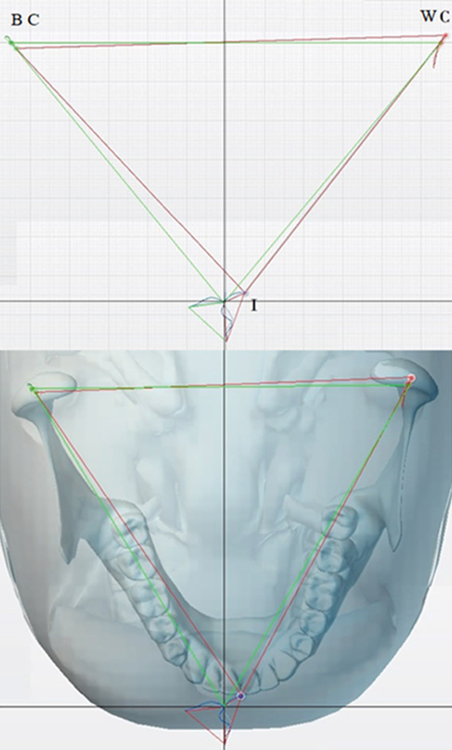
Fig. 1
Relationship between the gothic arch and Bonwill triangle.
I = Interincisive point.
C = Working condyle.
BC =Balancing condyle.
WC = Working condyle.
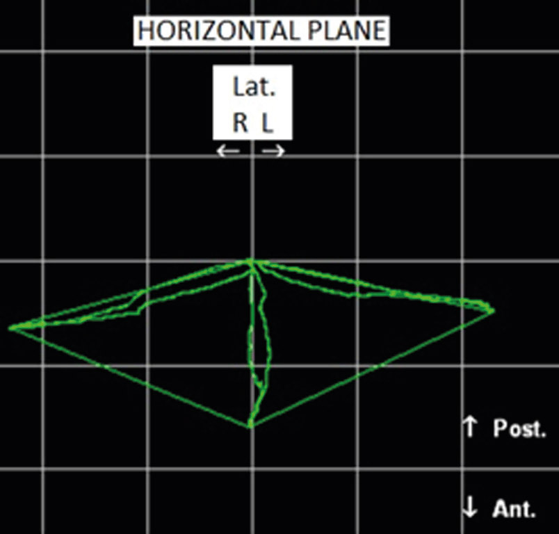
Fig. 2
Symmetrical gothic arch.
The following measurements of lateral excursion were taken with regard to tracing of left/right retrusion of the incisal point, tracing of left/right retrusion of the condylar point, tracing of left/right verticality of the incisal point and tracing of the left/right extent of the incisal point. We highlighted which of the two working condyle tracings appeared retrusive.
Results
Sixty-six clinical records of patients who had undergone axiographic examination were found and analyzed retrospectively. A total of 37 patients (25 females and 12 males) met the inclusion criteria and were included in the study. All of the patients showed TMJ symptoms and signs like clicking, pain during mastication, and limited movement and opening of the jaw.
All of the axiographic tracings studied showed an asymmetrical gothic arch with retrusive laterality in an absolute sense with respect to the frontal plane or with respect to the contralateral laterality. Thirty-seven axiographic tracings of laterality in 37 patients were studied.
The values of the terminal points of the interincisive lateral trajectories were compared on both sides with the values of the terminal points of the trajectories of the corresponding working condyles. In 81.08% of the cases, there was posteriorization of the working condyle on the side in which the interincisive point showed more retrusive laterality.
Discussion
This study was based on the analysis of lateral excursion, taking into consideration two gnathological concepts:
(1) The Bonwill triangle described in 1858 as an equilateral triangle placed on the horizontal plane, with the 3 vertices placed at the interincisive point of the jaw and in the middle of the two condyles. The sides of this triangle are 10 cm, although the correctness of this measurement has been questioned.15Christensen FT. The effect of Bonwill’s triangle on complete dentures. → J Prosthet Dent. 1959 Sep–Oct;9(5):791–6.
(2) The gothic arch that Gysi in 1930 first described with his recordings is a polygon created by the interincisive point on the horizontal plane with lateral and extreme protrusive movements.16Gysi A. Handbuch der Zahnheilkunde. Vol. 3. [Manual of dentistry]. Vol. 3. → Munich: J.F. Bergmann; 1930. 1007 p.
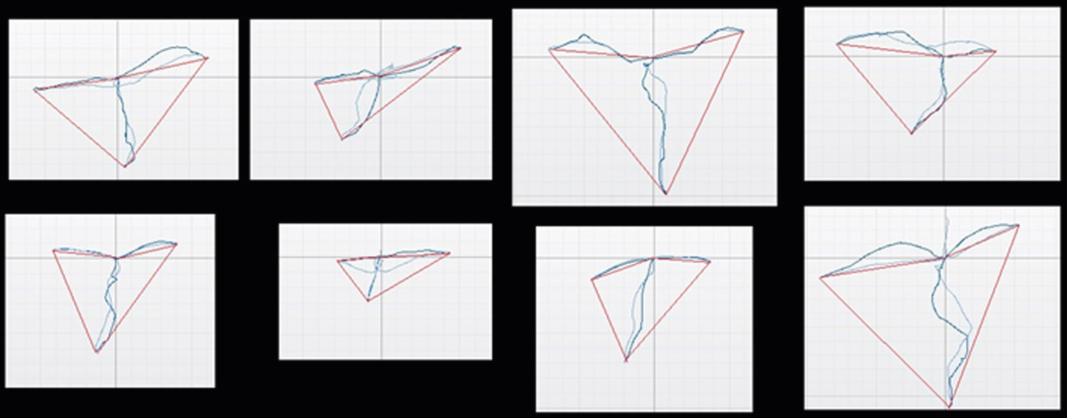
Asymmetrical gothic arch.
Keeping these two concepts in mind, in the present study, it was attempted to understand the relationship during lateral excursion on the horizontal plane between the shape of the gothic arch generated by the interincisive point (I) and the displacement of the vertex of the Bonwill triangle corresponding to the working condyle (C; Fig. 1). Although this may seem a very theoretical and abstract topic, the assessment of these relationships is important for the diagnosis and therapy of dysfunctional patients. In classical gnathology, the tracings of laterality on the horizontal plane are represented in a very regular and symmetrical way: a symmetrical frontal and lateral shift on both sides (Fig. 2). In kinesiographic tracings of dysfunctional patients, a deformation of the gothic arch is almost always observed, in which 1 or both tracings of laterality tend to lose the anterolateral direction, in order to acquire a tendency toward posteriority (Fig. 3).
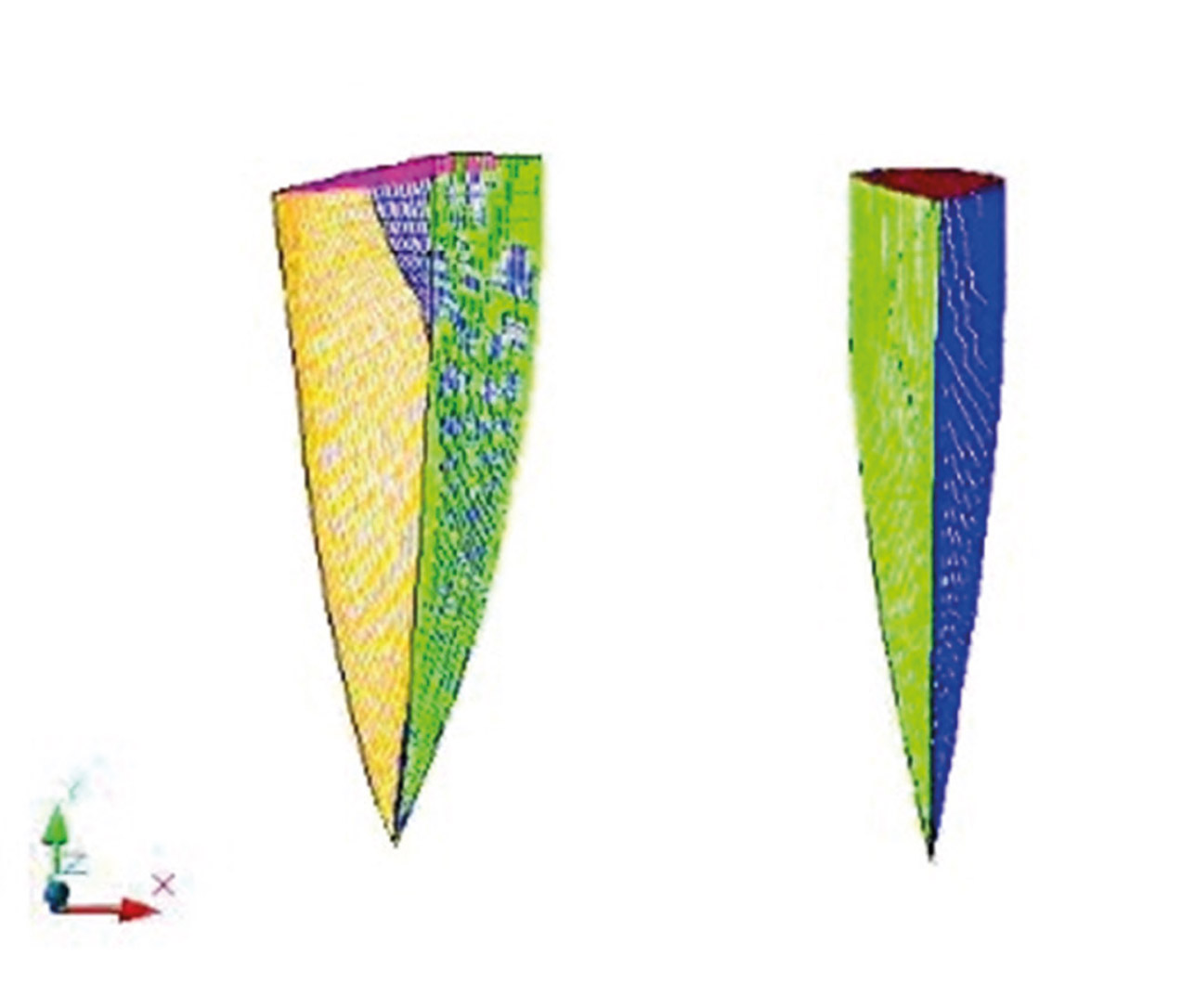
The gothic arch can be interpreted as a section on the horizontal plane of the volume identified by Posselt in his diagrams. This volume, formed by the extreme movements of lateral and protrusive opening, delimits the dynamic freedom that the jaw can express, during the functional movements that occur essentially within this perimeter.17Posselt U. Physiology of occlusion and rehabilitation. 2nd ed. → Oxford: Blackwell Scientific; 1968. 331 p. A large and symmetrical volume indicates a wide and symmetrical freedom of movement of the muscular structures of the jaw and of a physiological range of movement of the TMJ, essential in physiological alternating unilateral mastication (Fig. 4).
In 1984, Mongini put in geometrical relation the interincisive point with the condyles on the horizontal plane and in the static position of maximum intercuspation, statically describing the relations.18Mongini F. L’Apparato stomatognatico: funzione disfunzione e riabilitazione. Ed. internazionali. [The Stomatognathic apparatus: function dysfunction and rehabilitation.] → Milan: Scienza e tecnica dentistica; 1984. 372 p. With the kinesiographic layout of the laterality on the horizontal plane (gothic arch), the movement of the interincisive point can be kinematically put in relation to the movement effected by the working condyle: (1) If point B (balancing condyle) always moves in an anteromedial direction. (2) if point L (working condyle) has an anterolateral or posterolateral progression, then (3) point I will have the same trend of movement as point L (Fig. 5).
In a previous study, a relationship was established between the trajectories of the movement of the interincisive point on the frontal plane (PFMA) with those described by the same point on the horizontal plane (HMFA), highlighting that the steeper laterality on the frontal plane tends to be retrusive on the horizontal plane.19Papini A, Cesaretti G, De Fabianis P. Kinesiographic analysis of lateral excursive movement on the horizontal plane: the retrusive component. → J Oral Science Rehabilitation. 2017 Mar;3(1):60–70. In the present study, it was attempted to outline how the tracing of retrusive laterality of the interincisive point tends to correspond to posterior displacement of the apex of the Bonwill triangle of the working condyle.
The posteriorization of the condyle from its physiological position in the mandibular fossa, tends to create a compression of the soft retrodiscal tissue against the posterior wall of the mandibular fossa. The bilaminar zone of the retrodiscal tissue, being rich in vessels, nerve endings, adipose tissue and elastic fibers, is histologically unsuitable to support compressive forces.
-
-
Fig. 4
Different shape and size Posselt volume.
-

-
Fig. 5
Relationship between the interincisive point and working condyle.
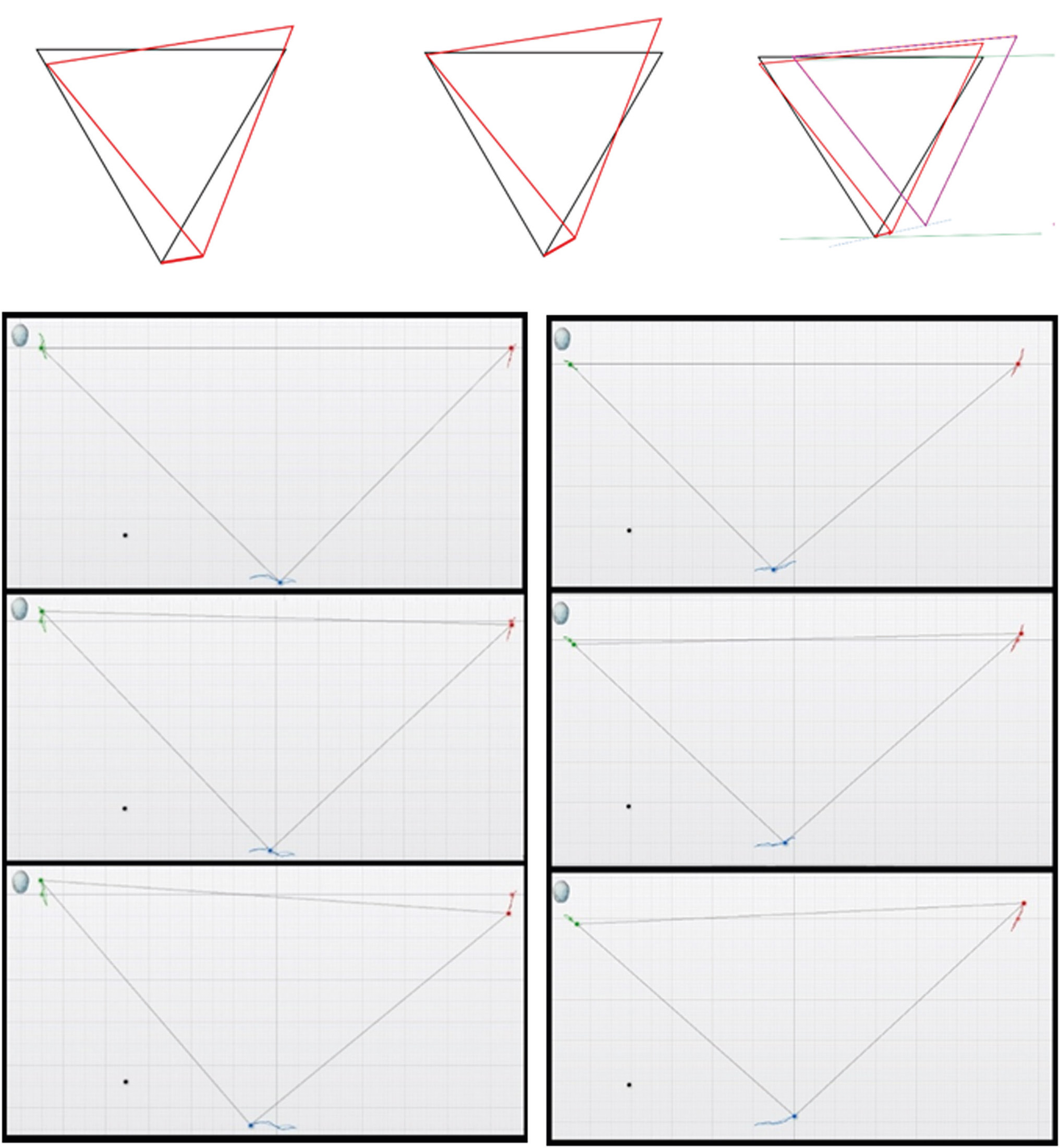
Relationship between the movement of the interincisive point and retrusion of the working condyle.
When subjected to repeated traumatic compressions, this soft structure reacts with inflammation that might result in fibrotic tissue composed of loose connective tissue,20Mazza D, Stasolla A, Kharrub Z, Maccioni F, Marini M. Valutazione con RM delle alterazioni morfo-strutturali del tessuto retrodiscale nell’incoordinazione condilo-discale dell’ATM: utilità delle sequenze TSE T2 pesate individualizzate. [MRI evaluation of morpho-structural alterations of the retrodiscal tissue in condylomeniscal incoordination of the TMJ: usefulness of individualised T2-weighted TSE sequences]. → Radiol Med. 2004;107:261–8. Italian.,21Yang L, Wang H, Wang M, Ohta Y, Suwa F. Development of collagen fibers and vasculature of the fetal TMJ. → Okajimas Folia Anat Jpn. 1992 Oct;69(4):145–55. 21. Pereira FJ, Lundh H, Eriksson L, Westesson PL. Microscopic changes in the retrodiscal tissues of painful temporomandibular joints. → J Oral Maxillofac Surg. 1996 Apr;54(4):461–8; discussion 469. with a significant increase in fibroblast density, the presence of restricted and obliterated artery lumina and a significantly lower distribution of elastic fibers.22Pereira FJ, Lundh H, Eriksson L, Westesson PL. Microscopic changes in the retrodiscal tissues of painful temporomandibular joints. → J Oral Maxillofac Surg. 1996 Apr;54(4):461–8; discussion 469.
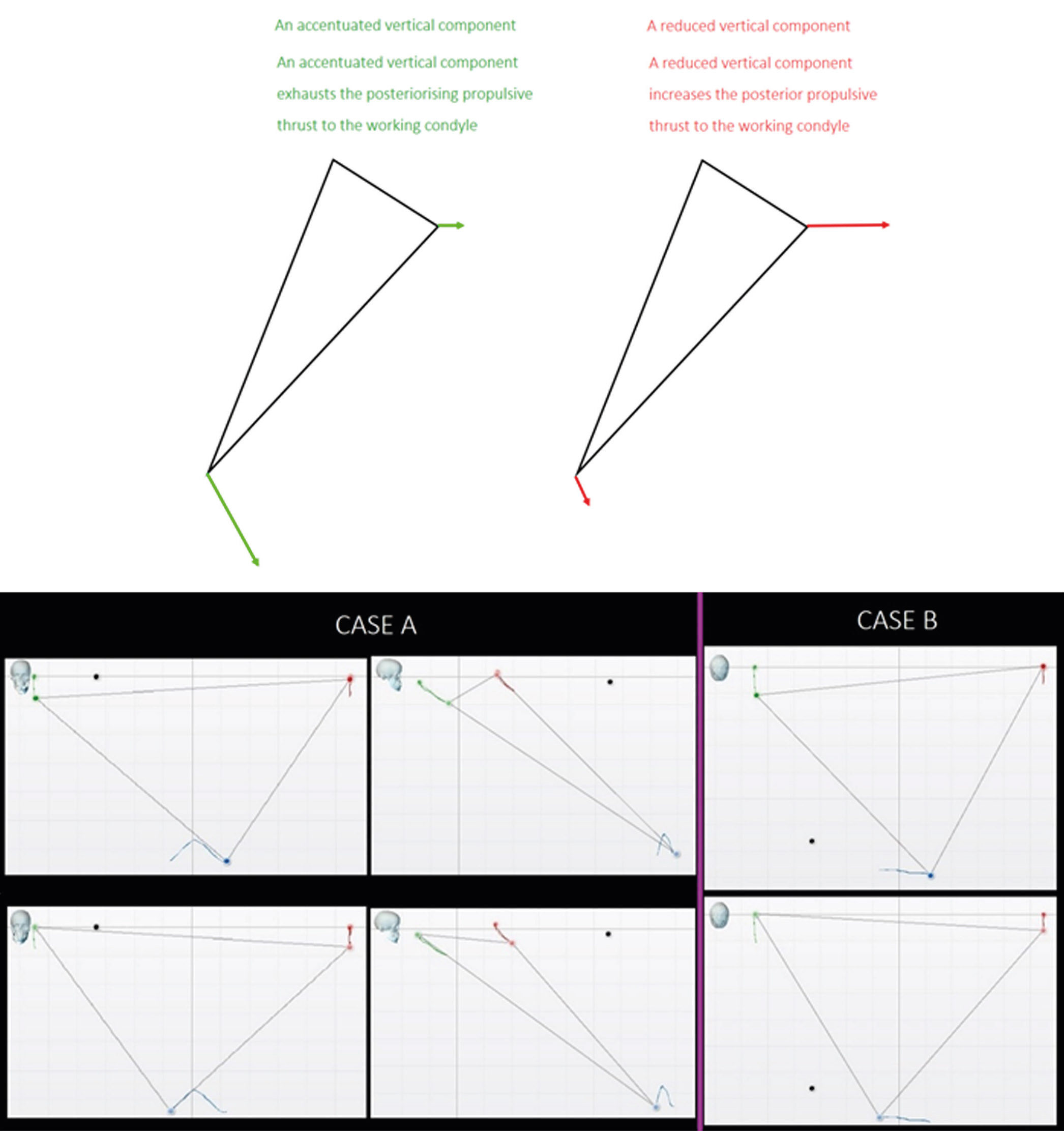
The retrusive thrust is attenuated by the vertical component of the lateral excursion.
During the phase of the power stroke in the crushing of the bolus, a functional moment in which the most intense forces of mastication develop, condylar retrusion tends to make the mastication traumatic, and this tends to induce mastication on the opposite side. In all of the tracings of laterality with retrusion of the interincisive point, there was a tendency to condylar retrusion. If in the lateral excursion the interincisive point presents an absolute retrusive trajectory, or relative to the contralateral movement, the corresponding working condyle tends to show a tendency to retrusion.
It must be remembered that in retrusive laterality of the interincisive point, condylar retrusion cannot be determined in a strictly geometric way. In fact, the posteriorization of the condyle will be influenced by other factors and will appear to be (1) directly proportional to both the posteriorization of the laterality tracing and its extension (Fig. 6), or (2) inversely proportional to the verticality of the movement of the interincisive point on the frontal plane. The lateral excursion is expressed on the 3 planes of the space, and the vertical component tends to exhaust the retrusive thrust of the working condyle (Fig. 7).
A steep PFMA on the frontal plane tends to correspond to a retrusive HMFA on the horizontal plane; a retrusive HMFA tends to induce retrusion of the corresponding working condyle. The lateral tracings, essentially overlapping the guidance of the entry phase of the masticatory cycle, are indexes of masticatory function of that side, being important proprioceptive parameters on which both masticatory cycles and their physiological alternation are set. Occlusal interference inducing retrusive laterality during mastication, that is, retrusion of the entry phase of the potentially compressive masticatory cycle of the delicate retrodiscal tissue, will make the system tend toward prevalent mastication on the opposite side.
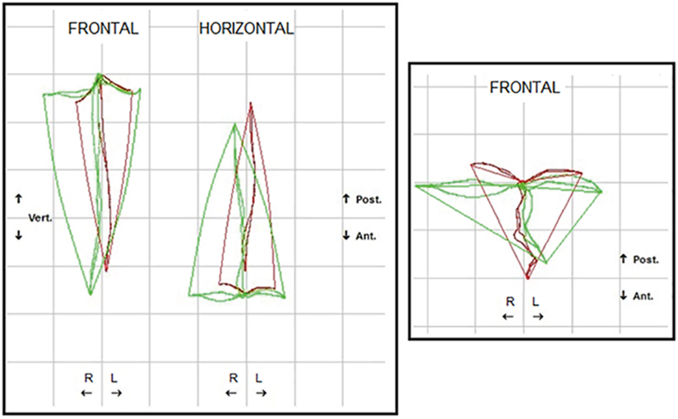
Change of Posselt volume after PFMA symmetrization.
The elimination of this interference by removing the functional obstacle through an accurate occlusal check will tend to favor a physiologically alternating mastication, resulting in symmetrization and enlargement of the gothic arch and with it of the entire Posselt volume, an indication of a better expression of muscular and articular function (Fig. 8).
The retrospective nature of the present study is the main limitation of the present report.
Competing interests
The present study was self-funded by the authors and their institutions. There is thus no conflict of interest to be declared.
Interview
Andrea Papini
Why did you conduct the research reported on in this paper?
For what reasons could others cite your paper?
How could your study’s findings have an impact on dentistry?
What is the relevance of your study’s findings to the daily practice of a dentist?
What are your recommendations for further investigation of the topic of your article?
References
| 1. | ↑ | Paglarelli Bergqvist L. The role of teeth in mammal history. → Braz J Oral Sciences. 2003 Jul–Sep;2(6):249–57. |
| 2. | ↑ | Kermack KA. The origin of mammals and the evolution of the temporomandibular joint. → Proc R Soc Med. 1972 Apr;65(4):389–92. |
| 3. | ↑ | Fritz J, Hummel J, Kienzle E, Streich WJ, Clauss M. To chew or not to chew: fecal particle size in herbivorous reptiles and mammals. → J Exp Zool A Ecol Genet Physiol. 2010 Nov 1;313(9):579–86. |
| 4. | ↑ | Herring SW. Functional morphology of mammalian mastication. → Am Zool. 1993;33(3):289–99. |
| 5. | ↑ | Lumsden AG, Osborn JW. The evolution of chewing: a dentist’s view of palaeontology. → J Dent. 1977 Dec;5(4):269–87. |
| 6. | ↑ | Inoue T. [Neural mechanisms of mastication]. → Brain Nerve. 2015 Feb;67(2):141–56. Japanese. |
| 7. | ↑ | Murdock DJ, Dong XP, Repetski JE, Marone F, Stampanoni M, Donoghue PC. The origin of conodonts and of vertebrate mineralized skeletons. → Nature. 2013 Oct 24;502(7472):546–9. |
| 8. | ↑ | Geerling E, Langenbach J, Van Eijden TM. Mammalian feeding motor patterns. → Am Zool. 2001 Dec;41(6):1338–51. |
| 9. | ↑ | Grossnickle DM. The evolutionary origin of jaw yaw in mammals. → Sci Rep. 2017 Mar 21;7:45094. |
| 10. | ↑ | Kay RF, Hiiemae KM. Jaw movement and tooth use in recent and fossil primates. → Am J Phys Anthropol. 1974 Mar;40(2):227–56. |
| 11. | ↑ | Williams SH, Vinyard CJ, Wall CE, Doherty AH, Crompton AW, Hylander WL. A preliminary analysis of correlated evolution in mammalian chewing motor patterns. integrative and comparative biology. → Integr Comp Biol. 2011 Aug;51(2):247–59. |
| 12. | ↑ | Gans C, De Vree F, Gorniak GC. Analysis of mammalian masticatory mechanisms: progress and problems. → Anat Histol Embryol. 1978 Sep;7(3):226–44. |
| 13. | ↑ | Planas P. Rehabilitación neuro-oclusal (RNO). [Neuro-occlusal rehabilitation]. → Barcelona: Salvat Editores; 1987. 317 p. |
| 14, 19. | ↑ | Papini A, Cesaretti G, De Fabianis P. Kinesiographic analysis of lateral excursive movement on the horizontal plane: the retrusive component. → J Oral Science Rehabilitation. 2017 Mar;3(1):60–70. |
| 15. | ↑ | Christensen FT. The effect of Bonwill’s triangle on complete dentures. → J Prosthet Dent. 1959 Sep–Oct;9(5):791–6. |
| 16. | ↑ | Gysi A. Handbuch der Zahnheilkunde. Vol. 3. [Manual of dentistry]. Vol. 3. → Munich: J.F. Bergmann; 1930. 1007 p. |
| 17. | ↑ | Posselt U. Physiology of occlusion and rehabilitation. 2nd ed. → Oxford: Blackwell Scientific; 1968. 331 p. |
| 18. | ↑ | Mongini F. L’Apparato stomatognatico: funzione disfunzione e riabilitazione. Ed. internazionali. [The Stomatognathic apparatus: function dysfunction and rehabilitation.] → Milan: Scienza e tecnica dentistica; 1984. 372 p. |
| 20. | ↑ | Mazza D, Stasolla A, Kharrub Z, Maccioni F, Marini M. Valutazione con RM delle alterazioni morfo-strutturali del tessuto retrodiscale nell’incoordinazione condilo-discale dell’ATM: utilità delle sequenze TSE T2 pesate individualizzate. [MRI evaluation of morpho-structural alterations of the retrodiscal tissue in condylomeniscal incoordination of the TMJ: usefulness of individualised T2-weighted TSE sequences]. → Radiol Med. 2004;107:261–8. Italian. |
| 21. | ↑ | Yang L, Wang H, Wang M, Ohta Y, Suwa F. Development of collagen fibers and vasculature of the fetal TMJ. → Okajimas Folia Anat Jpn. 1992 Oct;69(4):145–55. 21. Pereira FJ, Lundh H, Eriksson L, Westesson PL. Microscopic changes in the retrodiscal tissues of painful temporomandibular joints. → J Oral Maxillofac Surg. 1996 Apr;54(4):461–8; discussion 469. |
| 22. | ↑ | Pereira FJ, Lundh H, Eriksson L, Westesson PL. Microscopic changes in the retrodiscal tissues of painful temporomandibular joints. → J Oral Maxillofac Surg. 1996 Apr;54(4):461–8; discussion 469. |


Leave a Reply
Be the First to Comment!